| Original Articles |
|


|
|
|
| Effects of regional cerebral blood flow perfusion on learning and memory function and its molecular mechanism in rats |
| Cunli Xu1, Wenhua Wu2, Lingbin Kong3 |
1 Rehabilitation Medicine Center, The First People's Hospital of Jining, Jining, China;
2 Department of Neurology, The Second People's Hospital of Jining, Jining, China;
3 Shandong Institute of Behavioral Medical Science, Jining Medical College, Jining, China |
|
|
|
|
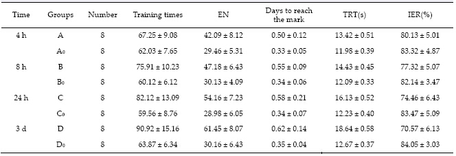
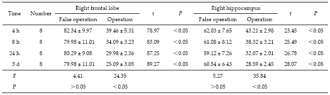
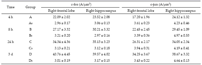
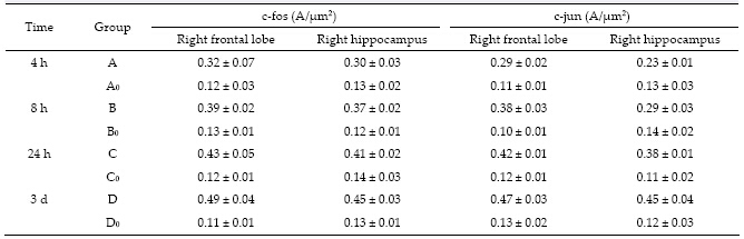
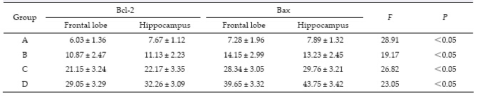
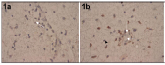
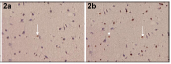
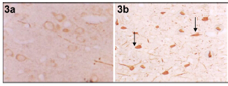
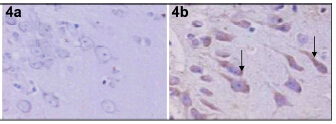
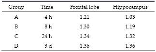
Abstract Objective: To study the effects of regional cerebral blood flow (rCBF) perfusion on learning and memory function in special brain areas and its molecular mechanism in rat. Methods: Sixty-four adult male healthy Sprague-Dawley (SD) rats were randomly divided into two groups: A false operation group and an operation group. The false operation group was randomly divided into four subgroups (A0, B0, C0, and D0) and the operation group was randomly divided into four subgroups (A, B, C, and D), with eight rats in each subgroup. The operation group underwent bilateral common carotid artery permanent ligation, while the other group only underwent a skin incision without the bilateral common carotid artery permanent ligation. Learning memory function of rats in each subgroup was measured using a Y-maze at 4 h, 8 h, 24 h, and 3 days after surgery. The rCBF in the right frontal lobe and hippocampus was detected using the Periflux PF model laser Doppler flowmetry and c-fos, c-jun, Bcl-2, and Bax protein expression in the right frontal lobe and hippocampus was measured using immunohistochemistry. Results: The rCBF in the right frontal lobule division and right hippocampus division was significantly lower in the operation group than in the false operation group (P < 0.05). The error number (EN), time to reach the target, and total reaction time (TRT) for the learning index using the Y-type labyrinth test in the operation group were significantly higher than that in the false operation group (P < 0.05); however, the active avoid rate in the operation group was significantly lower than that of the false operation group. Expression of c-fos and c-jun as well as the average absorbency in the right frontal lobule division and right hippocampus division in the operation group were significantly higher than those in the false operation group (P < 0.05). The number of Bax and Bcl-2-positive cells was significantly higher in the operation group, and the expression ratio of Bax/Bcl-2 in the operation group was significantly higher than that in the false operation group (P < 0.01). Conclusions: rCBF decrease can impair learning and memory function in rats, which may be related to the increased expression of c-fos, c-jun, Bcl-2, and Bax proteins in the t he frontal cortex and hippocampus.
|
|
Received: 11 January 2016
Published: 31 March 2016
|
|
Corresponding Authors:
Lingbin Kong, E-mail: klb3904@163.com
E-mail: klb3904@163.com
|
|
|
[1] Cao YG, Xu LJ, Xu L, Song ZK, Wu JM. Progress in the mechanism and prevention of skeletal muscle ischemiareperfusion injury. Med Recapitul 2009, 15(1): 126-129. (in Chinese)
[2] Séquier JM, Hunziker W, Richards G. Localization of calbindin D28 mRNA in rat tissues by in situ hybridization. Neurosci Lett 1988, 86(2): 155-160.
[3] Ryseck RP, Bravo R. c-JUN, JUN B, and JUN D differ in their binding affinities to AP-1 and CRE consensus sequences: Effect of FOS proteins. Oncogene 1991, 6(4): 533-542.
[4] Ohkubo Y, Arima M, Arguni E, Okada S, Yamashita K, Asari S, Obata S, Sakamoto A, Hatano M, O-Wang J, et al. A role for c-fos/Activator protein 1 in B lymphocyte terminal differentiation. J Immunol 2005, 174(12): 7703-7710.
[5] Han J, Guo HZ. Effect of ischemic preconditioning on expression of hsp70 and fos after focal cerebral infarction in rat. Chin J Clin Neurosci 2004, 12(2): 136-139. (in Chinese)
[6] Wiedmann R, Rosahl SK, Brinker T, Samii M, Nakamura M. Effect of acute and chronic bilateral visual deafferentation on c-fos immunoreactivity in the visual system of adult rats. Exp Brain Res 2013, 229(4): 595-607
[7] Montag-Sallaz M, Buonviso N. Altered odor-induced expression of c-fos and arg 3.1 immediate early genes in the olfactory system after familiarization with an odor. J Neurobiol 2002, 52(1): 61-72.
[8] Zhou F, Guo JC, Yang R, Gu J, Jin HB, Wu GC, Cheng JS. Effects of taurine on cerebral blood flow perfusion, cell apoptosis, and infarct volume in acute cerebral ischemic rats. In Taurine 6. Oja SS, Saransaari P, Eds. US: Springer, 2006, 583: 353-358.
[9] Blasiak A, Siwiec M, Grabowiecka A, Blasiak T, Czerw A, Blasiak E, Kania A, Rajfur Z, Lewandowski MH, Gundlach AL. Excitatory orexinergic innervation of rat nucleus incertuse—Implications for ascending arousal, motivation and feeding control. Neuropharmacology 2015, 99: 432-447.
[10] Herrera DG, Robertson HA. Activation of c-fos in the brain. Progr Neurobiol 1996, 50(2-3): 83-107.
[11] Herdegen T, Waetzig V. AP-1 proteins in the adult brain: Facts and fiction about effectors of neuroprotection and neurodegeneration. Oncogene 2001, 20(19): 2424-2437.
[12] Runyan JD, Dash PK. Distinct prefrontal molecular mechanisms for information storage lasting seconds versus minutes. Learn Mem 2005, 12(3): 232-238.
[13] Hu JL, Xie H, Long ZJ, Meng Q, Zhang YH, He YS. Effects of Ning-Shen Wen-Dan decoction to the expression of apoptosis protein Bax, Bcl-2 in hippocampus of rats. Chin J Basic Med Tradit Chin Med 2013, 19(4): 394-396. (in Chinese)
[14] Van Der Borght K, Wallinga AE, Luiten PGM, Eggen BJL, Van Der Zee EA. Morris water maze learning in two rat strains increases the expression of the polysialylated form of the neural cell adhesion molecule in the dentate gyrus but has no effect on hippocampal neurogenesis. Behav Neurosci 2005, 119(4): 926-932. |
|
Viewed |
|
|
|
Full text
|
|
|
|
|
Abstract
|
|
|
|
|
Cited |
|
|
|
|
| |
Shared |
|
|
|
|
| |
Discussed |
|
|
|
|