| Original Articles |
|


|
|
|
| Repairing skull defects in children with nano-hap/collagen composites: A clinical report of thirteen cases |
| Tuoyu Chen1, Yuqi Zhang1, Huancong Zuo1, Yapeng Zhao3, Chaoqiang Xue1, Bin Luo1, Qinglin Zhang1, Jin Zhu1, Xiumei Wang2, Fuzhai Cui2 |
1 Department of Neurosurgery, Tsinghua University Yuquan Hospital, Beijing 100040, China;
2 School of Materials Science and Engineering, Tsinghua University, Beijing 100084, China;
3 The Medical Center, Tsinghua University, Beijing 100084, China |
|
|
|
|
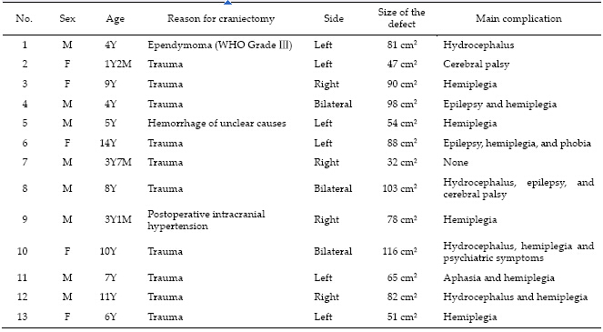
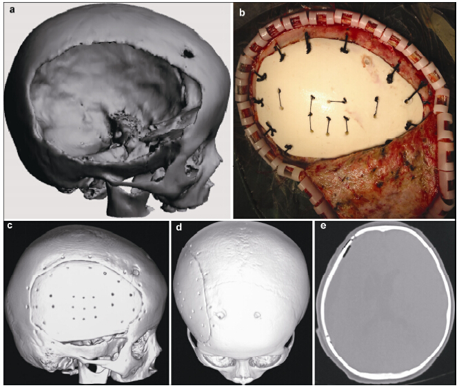
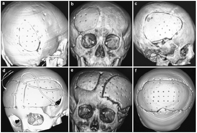
Abstract Objective: To evaluate the clinical results of repairing skull defects with biomimetic bone (nano-hap/collagen composites, NHACs) in children. Methods: Thirteen children with skull defects were treated with NHACs in our hospital. The NHACs molded with the help of a 3D printer were used in the operations. Results: All 13 operations were successful, and patients recovered without infection. Only one patient suffered from subcutaneous hydrops post-operation. The implanted NHACs remained fixed well after 1 year, and their CT HU values raised gradually. Skull shapes of children developed normally. Recovery of neurological and cognitive function was significant. Conclusions: NHAC, chosen to repair skull defects in children, can coexist with normal skull and reduce the negative effects on growth and development. NHAC could be a good choice for children with skull defects.
|
|
Received: 05 January 2016
Published: 31 March 2016
|
|
Corresponding Authors:
Yuqi Zhang, E-mail: yuqi9597@sina.com
E-mail: yuqi9597@sina.com
|
|
Cite this article:
Tuoyu Chen, Yuqi Zhang, Huancong Zuo, Yapeng Zhao, Chaoqiang Xue, Bin Luo, Qinglin Zhang, Jin Zhu, Xiumei Wang, Fuzhai Cui. Repairing skull defects in children with nano-hap/collagen composites: A clinical report of thirteen cases. Translational Neuroscience and Clinics, 2016, 2(1): 31-37.
URL:
http://tnc.tsinghuajournals.com/10.18679/CN11-6030/R.2016.005 OR http://tnc.tsinghuajournals.com/Y2016/V2/I1/31
|
|
|
[1] Josan VA, Sgouros S, Walsh AR, Dover MS, Nishikawa H, Hockley AD. Cranioplasty in children. Child's Nerv Syst 2005, 21(3): 200-204.
[2] Blum KS, Schneider SJ, Rosenthal AD. Methyl methacrylate cranioplasty in children: long-term results. Pediatr Neurosurg 1997, 26(1): 33-35.
[3] Takumi I, Akimoto M. Catcher's mask cranioplasty for extensive cranial defects in children with an open head trauma: A novel application of partial cranioplasty. Child's Nerv Syst 2008, 24(8): 927-932.
[4] Shah AM, Jung H, Skirboll S. Materials used in cranioplasty: A history and analysis. Neurosurgical Focus 2014, 36(4): E19.
[5] Edwards MSB, Oustehout DK. Autogeneic skull bone grafts to reconstruct large or complex skull defects in children and adolescents. Neurosurgery 1987, 20(2): 273-280.
[6] Qiu ZY, Zhang YQ, Zhang ZQ, Song TX, Cui FZ. Biodegradable mineralized collagen plug for the reconstruction of craniotomy burr-holes: A report of three cases. Transl Neurosci Clini 2015, 1(1): 3-9.
[7] Song Q, Hu K, Cui FZ, He ZY. Effect of high temperature on morphology and structure of nano-hydroxyapatite/collagen composite. Mater Sci Forum 2009, 610-613: 1360-1363.
[8] Itokawa H, Hiraide T, Moriya M, Fujimoto M, Nagashima G, Suzuki R, Fujimoto T. A 12 month in vivo study on the response of bone to a hydroxyapatite-polymethylmethacrylate cranioplasty composite. Biomaterials 2007, 28(33): 4922-4927.
[9] Du C, Cui FZ, zhang w, Feng QL, Zhu XD, de Groot K. Formation of calcium phosphate/collagen composites through mineralization of collagen matrix. J Biomed Mat Res 2000, 50: 518-527.
[10] Qiu ZY, Cui Y, Tao CS, Zhang ZQ, Tang PF, Mao KY, Wang XM, Cui FZ. Mineralized collagen: Rationale, current status, and clinical applications. Materials 2015, 8: 4733-4750.
[11] Okumura M, Ohgushi H, Tamai S. Bonding osteogenesis in coralline hydroxyapatite combined with bone marrow cells. Biomaterials 1991, 12(4): 411-416.
[12] Lian XJ, Qiu ZY, Wang CM, Guo WG, Zhang XJ, Dong YQ, Cui FZ. Structural and biomedical properties of zirconiahydroxyapatite nano-crystal ceramics. Biomater Tissue Eng 2013, 3(3): 330-334.
[13] Maire E, Withers PJ. Quantitative X-ray tomography. Int Mater Rev 2014, 59(1): 1-43. |
|
Viewed |
|
|
|
Full text
|
|
|
|
|
Abstract
|
|
|
|
|
Cited |
|
|
|
|
| |
Shared |
|
|
|
|
| |
Discussed |
|
|
|
|