| Short Communications |
|


|
|
|
| A new drainage tube device |
| Chao He, Ming Zhao, Dong Yang, Tianya Wu, Leiyu Qiu |
| The People's Hospital of Zhuji, Zhuji 311800, China |
|
|
|
|
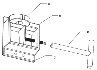
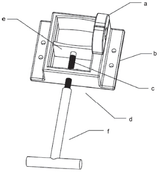
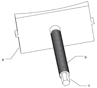
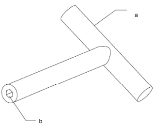
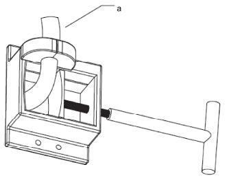
Abstract Objective: Drainage tubes (e.g., intracranial, abdominal cavity and thoracic) are commonly used to drain blood and fluid collections after surgery. It usually fails due to the lack of fixation perpendicular to the skin and the variety of tube materials. The objective of the article is to describe a new drainage tube device. Methods: This new device consists of a drainage tube fixator and diverter with a rotating handle. The fixator and diverter are fixed to the skin with a pair of wings, and the base comprises acircular arc with chamfering of the edge. The removable stopper and plastic diverter have a memory function. There are 2 holes on either side of the wings, by which they can be sewn to the skin or stapled for strength and stability. Results: The removable limiting stopper and plastic diverter with memory function work together to ensure that the drainage tube is firmly fixed. Therefore, the tube will not move either vertically or horizontally on the skin. Moreover, the device is fit for drainage tubes with different specifications and materials. Conclusions: The limiting stopper can be moved easily, which is conducive to local skin disinfection around the drainage tube. The device deserves clinical promotion.
|
|
Received: 30 December 2015
Published: 31 March 2016
|
|
Corresponding Authors:
Chao He, E-mail: zj.hechao@163.com
E-mail: zj.hechao@163.com
|
|
|
[1] Srinivasan VM, O'Neill BR, Jho D, Whiting DM, Oh MY. The history of external ventricular drainage. J Neurosurg 2014, 120(1): 228-236.
[2] Sun CR, Du HG, Yin LC, He M, Tian Y, Li HY. Choice for the removal of bloody cerebrospinal fluid in postcoiling aneurysmal subarachnoid hemorrhage: External ventricular drainage or lumbar drainage? Turk Neurosurg 2014, 24(5): 737-744.
[3] De Andrade AF, Paiva WS, Neville IS, Noleto GS, Junior AA, Sandon LHD, Bor-Seng-Shu E, Amorim RL, Teixeira MJ. Monoblock external ventricular drainage system in the treatment of patients with acute hydrocephalus: A pilot study. Med Sci Monit 2014, 20: 227-232.
[4] Kirmani AR, Sarmast AH, Bhat AR. Role of external ventricular drainage in the management of intraventricular hemorrhage; its complications and management. Surg Neurol Int 2015, 6: 188.
[5] Wang X, Dong Y, Qi XQ, Li YM, Huang CG, Hou LJ. Clinical review: Efficacy of antimicrobial-impregnated catheters in external ventricular drainage—A systematic review and meta-analysis. Crit Care 2013, 17(4): 234.
[6] Wiegand J, Hickson L, Merz TM. Indicators of external ventricular drainage-related infections—A retrospective observational study. Acta Neurochir 2016, 158(3): 595-601. |
|
Viewed |
|
|
|
Full text
|
|
|
|
|
Abstract
|
|
|
|
|
Cited |
|
|
|
|
| |
Shared |
|
|
|
|
| |
Discussed |
|
|
|
|