| Research Article |
|


|
|
|
| Controlled release of nerve growth factor and basic fibroblast growth factor combined with small-gap anastomosis enhances sciatic nerve regeneration |
| Xiyuan Wang1, Lin Chen2, Huancong Zuo2, Huagang Liu2, Liu Ji2, Shanker Sharma Hari3, Sharma Aruna3, Qiang Ao1,2 |
1 Department of Tissue Engineering, China Medical University, Shen Yang 110122, China;
2 Department of Neurosurgery, Tsinghua University Yuquan Hospital, Beijing 100040, China;
3 Laboratory of Cerebrovascular Research, Department of Surgical Sciences Anaesthesiology and Intensive Care Medicine, Uppsala University Hospital, Uppsala University, Se-75185 Uppsala, Sweden |
|
|
|
|
| Guide |
|
Abstract Objectives: Nerve regeneration after peripheral nerve injury is a slow process with a limited degree of functional recovery, resulting in a high disability rate. Thus, accelerating the rate of nerve regeneration and improving the degree of nerve repair is a clinical challenge. This study aimed to investigate the role of growth factor gel combined with small-gap nerve anastomosis in the regeneration of sciatic nerve injury in rats. This was achieved by injecting nerve growth factor (NGF) and basic fibroblast growth factor (bFGF) gel into a silicon chamber that bridged the transection of the nerve.
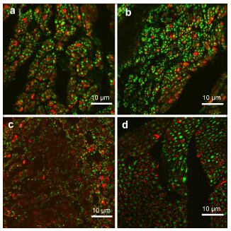
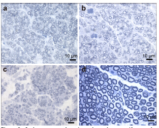
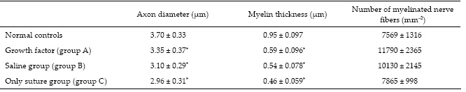
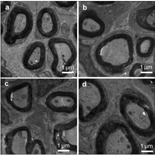
Methods: In 27 randomly chosen Sprague Dawley rats, a sharp blade was used to transect the right hind leg sciatic nerve. The rats were divided into 3 groups: in groups A and B, silicon tubes containing NGF and bFGF gel or saline, respectively, were used to bridge the nerve proximal and distal ends (3-mm gap), and in group C, the nerve proximal and distal ends were directly sutured. Eight weeks after surgery, the sciatic nerve function index, neural electrophysiology, and muscle wet weight as well as histological, ultrastructural, and immunohistochemical parameters were evaluated.
Results: The sciatic nerve function index, nerve conduction velocity, muscle wet weight, density of regenerated nerve fibers, and myelination in group A were better than those in group B or C, but the sciatic nerve function index, muscle wet weight, and thickness of myelination in the 3 groups were not significantly different (P > 0.05). There were no significant differences innerve conduction velocity between groups A and B (P > 0.05), but it was higher in both groups than that of group C (P < 0.05). The regenerated nerve fiber density in the 3 groups showed significant differences (P < 0.05).
Conclusions: Small-gap nerve anastomosis can provide a good regenerative microenvironment for rat sciatic nerve regeneration, and the combined strategy of growth factor gel with small-gap nerve anastomosis appears to have a superior effect on nerve repair.
|
|
Received: 20 March 2015
Published: 01 September 2015
|
|
|
| Fund: This work was supported by the National High Technology Research and Development Program of China (863 Program, No. 2012AA020905), and the Chow Tai Fook Medical Research Special Fund (No. 202836019-03). |
|
Corresponding Authors:
Qiang Ao, E-mail: aoqiang@tsinghua.edu.cn;Lin Chen, E-mail: chenlin_china@163.com
E-mail: aoqiang@tsinghua.edu.cn;chenlin_china@163.com
|
|
|
[1] Catrina S, Gander B, Madduri S. Nerve conduit scaffolds for discrete delivery of two neurotrophic factors. Eur J Pharm Biopharm 2013, 85(1): 139-142.
[2] Zhang ZJ, Kou YH, Yin XF, Wang YH, Zhang PX, Jiang BG. The effect of a small gap sleeve suture at the distal anastomosis of a nerve graft on promoting nerve regeneration and functional recovery. Artif Cells Nanomed Biotechnol 2013, 41(4): 282-288.
[3] Meek MF, Den Dunnen WFA, Schakenraad JM, Robinson PH. Long-term evaluation of functional nerve recovery after reconstruction with a thin-walled biodegradable poly (DLlactide-epsilon-caprolactone) nerve guide, using walking track analysis and electrostimulation tests. Microsurgery 1999, 19(5): 247-253.
[4] Butí M, Verdú E, Labrador RO, Vilches JJ, Forés J, NavarroX. Influence of physical parameters of nerve chambers on peripheral nerve regeneration and reinnervation. Exp Neurol 1996, 137(1): 26-33.
[5] Zhao LG, Yao KD. Advances in nerve regeneration through nerve conduit. BME & Clin Med 2003, 7(2): 120-123. (in Chinese)
[6] Lundborg G. Nerve regenration and repair. A review. Acta Orthop Scand 1987, 58(2): 145-169.
[7] Song XZ, Gu YD, Xu JG, Li JF. Comparison of suture and conduit nerve repair in different alignment situation. Chin J Hand Surg 1998, 14(4): 250-251. (in Chinese)
[8] Ahmed TAE, Dare EV, Hincke M. Fibrin: A versatile scaffold for tissue engineering applications. Tissue Eng Part B 2008, 14(2): 199-215.
[9] Ishii I, Mizuta H, Sei A, Hirose J, Kudo S, Hiraki Y. Healing of full-thickness defects of the articular cartilage in rabbits using fibroblast growth factor-2 and a fibrin sealant. J Bone Joint Surg Br 2007, 89(5): 693-700.
[10] Jeon O, Ryu SH, Chung JH, Kim SB. Control of basic fibroblast growth factor release from fibrin gel with heparin and concentrations of fibrinogen and thrombin. J Control Release 2005, 105(3): 249-259.
[11] Ho YC, Mi FL, Sung HW, Kuo PL. Heparin-functionalized chitosan-alginate scaffolds for controlled release of growth factor. Int J Pharm 2009, 376(1-2): 69-75.
[12] Tanihara M, Suzuki Y, Yamamoto E, Noguchi A, Mizushima Y. Sustained release of basic fibroblast growth factor and angiogenesis in a novel covalently crosslinked gel of heparin and alginate. Biomed Mater Res 2001, 56(2): 216-221. |
|
Viewed |
|
|
|
Full text
|
|
|
|
|
Abstract
|
|
|
|
|
Cited |
|
|
|
|
| |
Shared |
|
|
|
|
| |
Discussed |
|
|
|
|