| Short Communication |
|

|
|
|
| A newly developed open-end intracranial hematoma drainage tube |
| Chao He1,2, Nongnaphat Wanussakul1, Dong Yang1, Tianya Wu1 |
1 The People's Hospital of Zhuji, Zhuji 311800, China;
2 The Second Affiliated Hospital of Zhejiang, Hangzhou 310000, China |
|
|
|
|
| Guide |
|
Abstract Objective: To design a new open-end intracranial hematoma drainage tube for clinical application.
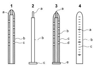
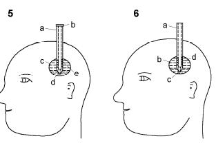
Methods: The newly developed device consists of two parts:the plunger and barrel. On one side, the barrel is bullet shaped with an opening tip. The plunger is located in the middle cavity of the tube barrel and extended out at the open-end. It was designed for strengthening the tube barrel and for convenience in performing the drainage procedure. It can be used by inserting the drainage tube into the lesion and pulling out the plunger, whereby blood will forcefully rise up inside the barrel, providing a satisfactory outcome. It is made for effusion drainage purposes. During the procedure, the drainage tip is placed at the deepest part of the intracranial hematoma to completely drain the blood. Moreover, the plunger fits tightly in the tube, preventing leakage during the operation. With the use of the device, brain can be separated. In addition, the device can help reduce the risk of cerebral damage because of the small operating area. The barrel sidewall has matching opening holes bilaterally and equally for exchanging substances between the inner and outer parts. The overlapping ratio in each horizontal pair is around 1/3-1/2. Each pair on the opposite side will form a different pressure. Thus, the opening holes will not easily get blocked with blood clot.
Results: Blood and accumulated liquid from the deepest part of the intracranial hematoma can be directly drawn through the drainage tube without damaging a large area. The tube does not get blocked easily and allows for complete removal of the hematoma.
Conclusions: The device is asuitable instrument for clinical application.
|
|
Received: 20 May 2016
Published: 30 June 2016
|
| Fund: Supported by the Zhejiang Province General Medical Health Research Program (No. 2016KYB315). |
|
Corresponding Authors:
Chao He,E-mail:zj.hechao@163.com
E-mail: zj.hechao@163.com
|
|
|
[1] Kalani MYS, Filippidis A, Martirosyan NL, Theodore N. Cerebral herniation as a complication of chest tube drainage of cerebrospinal fluid after injury to the spine. World Neurosurg 2013, 79(5-6):798.e17-798.e19.
[2] Wang X, Dong Y, Qi XQ, Li YM, Huang CG, Hou LJ.Clinical review:Efficacy of antimicrobial-impregnated catheters in external ventricular drainage-A systematic review and meta-analysis. Crit Care 2013, 17:234.
[3] Cinibulak Z, Aschoff A, Apedjinou A, Kaminsky J, Trost HA, Krauss JK. Current practice of external ventricular drainage:Asurvey among neurosurgical departments in Germany. Acta Neurochir 2016, 158(5):847-853.
[4] Feng Y, He JQ, Liu B, Yang LK, Wang YH. Endoscope-assisted keyhole technique for hypertensive cerebral hemorrhage in elderly patients:A randomized controlled study in 184 patients. Turk Neurosurg 2016, 26(1):84-89.
[5] Jamjoom AAB, Kolias AG, Zaben M, Chari A, Kitchen J, Joannides A, Brennan PM, Kandasamy J, Gatscher S, Gray WP, et al. External ventricular drainage:Is it time to look at national practice? Br J Neurosurg 2015, 29:9-10.
[6] Fisher CM. Hypertensive cerebral hemorrhage. Demonstration of the source of bleeding. J Neuropathol Exp Neurol 2003, 62(1):104-107.
[7] Tahir MZ, Sobani ZA, Murtaza M, Enam SA. Long-tunneled versus short-tunneled external ventricular drainage:Prospective experience from a developing country. Asian J Neurosurg 2016, 11(2):114-117.
[8] Aurangzeb A, Khan SA, Ahmed E, Mehmood S, Ali A, Zadran KK, Hussain S. Predisposing factors, clinical presentation and outcome of repeated aspiration in cerebral abscess through a drainage tube in situ. J Ayub Med Coll Abbottabad 2011, 23(4):58-60.
[9] Wang K, Du HG, Yin LC, He M, Hao BL, Chen L.Which side of lateral ventricles to choose during external ventricular drainage in patients with intraventricular hemorrhage:Ipsilateral or contralateral? J Surg Res 2013, 183(2):720-725.
[10] Kirmani A, Sarmast A, Bhat A. Role of external ventricular drainage in the management of intraventricular hemorrhage; its complications and management. Surg Neurol Int 2015, 6(1):188. |
|
Viewed |
|
|
|
Full text
|
|
|
|
|
Abstract
|
|
|
|
|
Cited |
|
|
|
|
| |
Shared |
|
|
|
|
| |
Discussed |
|
|
|
|