摘要 Objectives: To discuss the bleeding mechanisms after removing a medulla oblongata hemangioblastoma. Methods: A 42-year-old male patient was diagnosed with a medulla oblongata hemangioblastoma. Preoperative cranial magnetic resonance imaging, computed tomography angiography and post-surgery computed tomography were completed during clinical procedure. We also reviewed the related literatures. Results: The preoperative computed tomography angiography did not demonstrate any intracranial aneurysm. But, the patient had a fatal subarachnoid hemorrhage with ventricular hemorrhage 4 hours after surgery following the post-surgery computed tomography. Conclusions: Subarachnoid hemorrhage after surgery of the medulla oblongata hemangioblastoma is very rare. Delayed postoperative hemorrhage seems the most reasonable explanation of Subarachnoid hemorrhage in our case.
Abstract: Objectives: To discuss the bleeding mechanisms after removing a medulla oblongata hemangioblastoma. Methods: A 42-year-old male patient was diagnosed with a medulla oblongata hemangioblastoma. Preoperative cranial magnetic resonance imaging, computed tomography angiography and post-surgery computed tomography were completed during clinical procedure. We also reviewed the related literatures. Results: The preoperative computed tomography angiography did not demonstrate any intracranial aneurysm. But, the patient had a fatal subarachnoid hemorrhage with ventricular hemorrhage 4 hours after surgery following the post-surgery computed tomography. Conclusions: Subarachnoid hemorrhage after surgery of the medulla oblongata hemangioblastoma is very rare. Delayed postoperative hemorrhage seems the most reasonable explanation of Subarachnoid hemorrhage in our case.
Xiang Yang, Yuekang Zhang, Xuesong Liu, Maojun Chen. Subarachnoid hemorrhage after surgery of the medulla oblongata hemangioblastoma: A case report[J]. 临床转化神经科学, 2016, 2(3): 195-198.
Xiang Yang, Yuekang Zhang, Xuesong Liu, Maojun Chen. Subarachnoid hemorrhage after surgery of the medulla oblongata hemangioblastoma: A case report. Translational Neuroscience and Clinics, 2016, 2(3): 195-198.
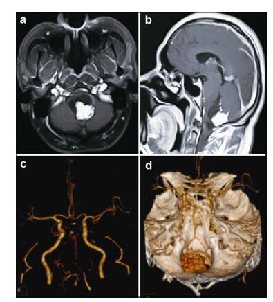
20161117201304 Figure 1 (a, b) Preoperative contrast-enhanced magnetic resonance images revealed a cystic solid hemangioblastoma, which was located on the dorsal part of the medulla oblongata. (c, d) Preoperative computed tomography angiography (CTA) images revealed that the bilateral vertebral arteries, basilar artery, and bilateral posterior cerebral arteries were slender and the right posterior cerebral artery was stenotic. It also showed the relationship between the tumor and the feeding arteries. No aneurysm was seen on the CTA scans
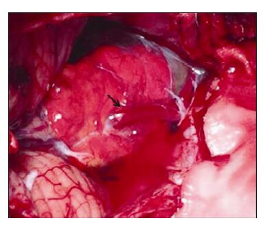
20161117201323 Figure 2 The arrow on the intraoperative photograph is pointing to a large draining vein of the tumor, and the tumor margins were exposed completely in this operative field.
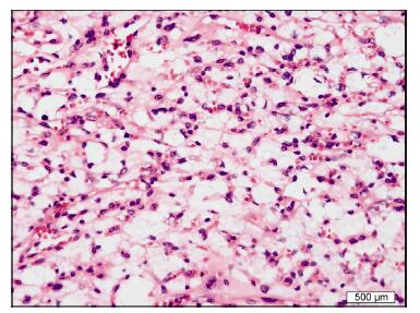
20161117201342 Figure 3 Histological examination showed round-to-oval nuclei and abundant clear cytoplasm in most cells, confirming a hemangioblastoma.
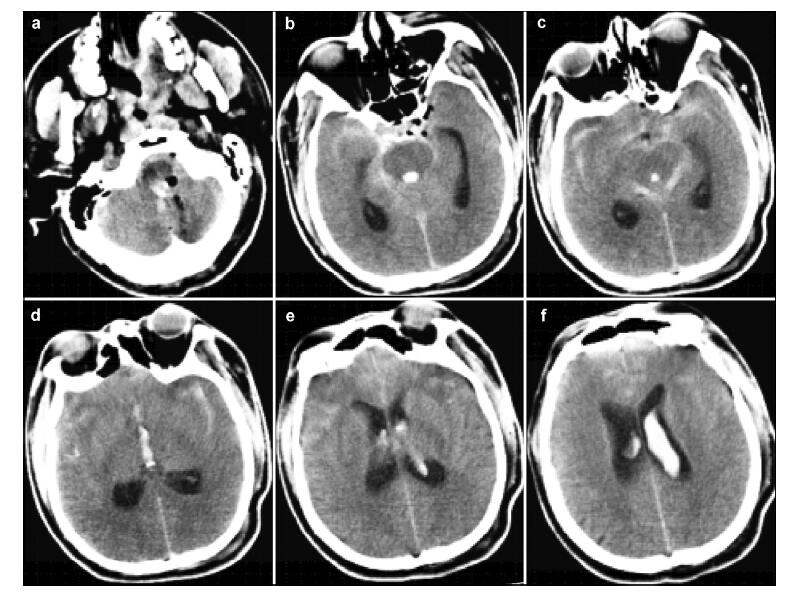
20161117201413 Figure 4 (a–f) Postoperative emergency CT scan showing extensive intracranial subarachnoid hemorrhage, and fourth and left lateral ventricular hemorrhage.
[1] Pavesi G, Berlucchi S, Munari M, Manara R, Scienza R, Opocher G. Clinical and surgical features of lower brain stem hemangioblastomas in von Hippel-Lindau disease. Acta Neurochir (Wien) 2010, 152(2):287-292.
[2] Gläsker S, Van Velthoven V. Risk of hemorrhage in heman-gioblastomas of the central nervous system. Neurosurgery 2005, 57(1):71-76.
[3] Fujii H, Higashi S, Hashimoto M, Shouin K, Hayase H, Kimura M, Yamamoto S. Hemangioblastoma presenting with fourth ventricular bleeding. Case report. Neurol Med Chir (Tokyo) 1987, 27(6):545-549.
[4] Ros de San Pedro J, Rodríguez FA, Ñíguez BF, Sánchez JFML, López-Guerrero AL, Murcia MF, Vilar AMRE. Massive hemorrhage in hemangioblastomas. Neurosurg Rev 2010, 33(1):11-26.
[5] Kallmes DF, Layton K, Marx WF, Tong F. Death by nondiagnosis:Why emergent CT angiography should not be done for patients with subarachnoid hemorrhage. Am J Neuroradiol 2007, 28(10):1837-1838.
[6] Sharma MS, Jha AN. Ruptured intracranial aneurysm associated with von Hippel-Lindau syndrome:A molecular link? J Neurosurg 2006, 104(2):90-93.
[7] Klingler JH, Krüger MT, Lemke JR, Jilg C, Van Velthoven V, Zentner J, Neumann HPH, Gläsker S. Sequence variations in the von Hippel-Lindau tumor suppressor gene in patients with intracranial aneurysms. J Stroke Cerebrovasc Dis 2013, 22(4):437-443.
[8] Chun YI, Cho J, Moon CT, Koh YC. Delayed fatal cerebellar hemorrhage caused by hemangioblastoma after successful radiosurgical treatment. Acta Neurochir (Wien) 2010, 152(9):1625-1627.
[9] Gekka M, Yamaguchi S, Kazumata K, Kobayashi H, Motegi H, Terasaka S, Houkin K. Hemorrhagic onset of hemangioblastoma located in the dorsal medulla oblongata presenting with tako-tsubo cardiomyopathy and neurogenic pulmonary edema:A case report. Case Rep Neurol 2014, 6(1):68-73.
 2016, Vol. 2