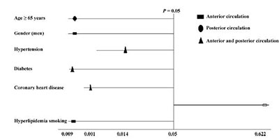
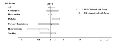
摘要 Objective: To discuss the correlation among intracranial arterial stenosis and its risk factors. Methods: A total of 486 patients with transient ischemic attack (TIA) or ischemic cerebral infarction were examined using color doppler flow imaging (CDFI) and transcranial doppler ultrosonography (TCD).According to the degrees of extracranial arterial stenosis,patients with mild-to-moderate extracranial stenosis were classified into group A (435 cases) while those with constant severe stenosis or occlusion were classified into group B (51 cases).The differences between the two groups of risk factors were compared,and the multi-factor logistic regression analysis of risk factors associated with moderately severe intracranial arterial stenosis was performed. Results: ① The risk factors that were significantly associated with intracranial arterial stenosis included age (P=0.034) and gender (P=0.044).② Intracranial artery stenosis was observed in both anterior and posterior cerebral arteries in patients with hypertension,diabetes,and coronary heart disease respectively (P< 0.05).③ Compared with group A,patients in group B were older (P=0.000),with a higher proportion of men (P=0.037),and the intracranial arterial stenosis degrees were significantly higher (P=0.013).④ Multi-factor logistic regression analysis showed that diabetes is a risk factor for moderately severe intracranial arterial stenosis (P< 0.05),and hyperlipidemia is a protective factor (P=0.012). Conclusions: Age,gender,hypertension,diabetes,coronary heart disease,and smoking are risk factors for the distribution of intracranial arterial stenosis.The degrees of intracranial arterial stenosis are related with extracranial arterial stenosis.Diabetes is a risk factor for moderately severe intracranial arterial stenosis while hyperlipidemia is a protective factor.
Abstract: Objective: To discuss the correlation among intracranial arterial stenosis and its risk factors. Methods: A total of 486 patients with transient ischemic attack (TIA) or ischemic cerebral infarction were examined using color doppler flow imaging (CDFI) and transcranial doppler ultrosonography (TCD).According to the degrees of extracranial arterial stenosis,patients with mild-to-moderate extracranial stenosis were classified into group A (435 cases) while those with constant severe stenosis or occlusion were classified into group B (51 cases).The differences between the two groups of risk factors were compared,and the multi-factor logistic regression analysis of risk factors associated with moderately severe intracranial arterial stenosis was performed. Results: ① The risk factors that were significantly associated with intracranial arterial stenosis included age (P=0.034) and gender (P=0.044).② Intracranial artery stenosis was observed in both anterior and posterior cerebral arteries in patients with hypertension,diabetes,and coronary heart disease respectively (P< 0.05).③ Compared with group A,patients in group B were older (P=0.000),with a higher proportion of men (P=0.037),and the intracranial arterial stenosis degrees were significantly higher (P=0.013).④ Multi-factor logistic regression analysis showed that diabetes is a risk factor for moderately severe intracranial arterial stenosis (P< 0.05),and hyperlipidemia is a protective factor (P=0.012). Conclusions: Age,gender,hypertension,diabetes,coronary heart disease,and smoking are risk factors for the distribution of intracranial arterial stenosis.The degrees of intracranial arterial stenosis are related with extracranial arterial stenosis.Diabetes is a risk factor for moderately severe intracranial arterial stenosis while hyperlipidemia is a protective factor.
20170712151214 Figure 1 Relationship among risk factors and the distribution of arterial stenosis.
20170712151222 Figure 2 Multi-factor logistic regression analysis of patients with moderately severe intracranial arterial stenosis. CI: Confidence intervals; OR: Odds Ratio.
[1]
Ois A, Gomis M, Rodríguez-Campello A, Cuadrado-Godia E, Jiménez-Conde J, Pont-Sunyer C, Cuccurella G, Roquer J. Factors associated with a high risk of recurrence in patients with transient ischemic attack or minor stroke. Stroke 2008, 39(6):1717-1721.
[2]
Wang YJ, Zhao XQ, Liu LP, Soo YOY, Pu YH, Pan YS, Wang YL, Zou XY, Leung TWH, Cai YF, et al. Prevalence and outcomes of symptomatic intracranial large artery stenoses and occlusions in China:The Chinese Intracranial Atherosclerosis (CICAS) Study. Stroke 2014, 45(3):663-669.
[3]
Accorsi F. Color Doppler of the extracranial and intracranial arteries in the acute phase of cerebral ischemia. J Ultrasound 2013, 16(4):187-193.
[4]
Turan TN, Makki AA, Tsappidi S, Cotsonis G, Lynn MJ, Cloft HJ, Chimowitz MI, the WASID Investigators. Risk factors associated with severity and location of intracranial arterial stenosis. Stroke 2010, 41(8):1636-1640.
[5]
Yamada S, Kobayashi M, Watanabe Y, Miyake H, Oshima M. Quantitative measurement of blood flow volume in the major intracranial arteries by using 123i-iodoamphetamine SPECT. Clin Nucl Med 2014, 39(10):868-873.
[6]
Holzer K, Sadikovic S, Esposito L, Bockelbrink A, Sander D, Hemmer B, Poppert H. Transcranial Doppler ultrasonography predicts cardiovascular events after TIA. BMC Med Imaging 2009, 9:13.
[7]
Wang XY, Chen L, Ao Q, Sharma A, Shanker Sharma H. Progress in the research and development of nerve conduits. Transl Neurosci Clin 2015, 1(2):97-101.
[8]
Guan JX, Zhou Q, Ouyang HQ, Zhang SF, Lu ZN. The diagnostic accuracy of TCD for intracranial arterial stenosis/occlusion in patients with acute ischemic stroke:The importance of time interval between detection of TCD and CTA. Neurol Res 2013, 35(9):930-936.
[9]
Wong KS, Huang YN, Gao S, Lam WWM, Chan YL, Kay R. Intracranial stenosis in Chinese patients with acute stroke. Neurology 1998, 50(3):812-813.
[10]
Teng MMH, Jen SL, Chiu FY, Kao YH, Lin CJ, Chang FC. Change in brain perfusion after extracranial-intracranial bypass surgery detected using the mean transit time of computed tomography perfusion. J Chin Med Assoc 2012, 75(12):649-653.
[11]
Suri MFK, Johnston SC. Epidemiology of intracranial stenosis. J Neuroimaging 2009, 19(Suppl 1):11S-16S.
[12]
Su LL, Huang HW, Tan SQ, Wu XH, Zhou GJ. Prevalence of asymptomatic intracranial artery stenosis in middle-aged and elderly population in the community of Foshan city, Guangdong province:A cross-sectional study. Chinese J Epidemiol 2011, 32(5):469-472.
[13]
Kang J, Kim N, Oh CW, Kwon OK, Jung CK, Kim WJ, Park JH, Ko Y, Noh WY, Jang MU, et al. Symptomatic steno-occlusion of cerebral arteries and subsequent ischemic events in patients with acute ischemic stroke. J Stroke Cerebrovasc Dis 2014, 23(5):e347-e353.
[14]
Kim BJ, Lee SH, Kang BS, Yoon BW, Roh JK. Diabetes increases large artery diseases, but not small artery diseases in the brain. J Neurol 2008, 255(8):1176-1181.
[15]
Lin T, Lin YH, Kao LL, Kao YH, Yang YH, Chou PS, Wu MN. An association between the location of white matter changes and the behavioral and psychological symptoms of dementia in Alzheimer's disease patients. Transl Neurosci Clin 2016, 2(1):8-16.
[16]
Mendes I, Baptista P, Soares F, Oliveira V, Ferro JM. Diabetes mellitus and intracranial stenosis. Rev Neurol 1999, 28(11):1030-1033.
[17]
Miyazawa N, Akiyama I, Yamagata Z. Analysis of incidence and risk factors for progression in patients with intracranial steno-occlusive lesions by serial magnetic resonance angiography. Clin Neurol Neurosurg 2007, 109(8):680-685.
[18]
Irace C, Pujia A, Motti C, Massimo F, Gnasso A. Carotid atherosclerosis in subjects with different hyperlipidaemia phenotypes. Int Angiol 1998, 17(1):15-21.
[19]
Shen Y, Wang J, Wu JW, Qu WK, Wang CX, Gao X, Zhou Y, Wang AX, Wu SL, Zhao XQ. Elevated plasma total cholesterol level is associated with the risk of asymptomatic intracranial arterial stenosis. PLoS One 2014, 9(7):e101232.
[20]
Xue MZ, Li YJ, Gao XG, Zhang CF. Atherosclerotic stenosis of intracranial and extracranial cerebral arteries in patients with cerebral infarction and the correlative factors. National Med J China 2011, 91(11):762-765. (in Chinese)
Wu JW, Zhang Q, Yang HJ, Gao X, Zhou Y, Wang AX, Wang CX, Zhang SF, Wu SL, Zhao XQ. Association between non-high-density-lipoprotein-cholesterol levels and the prevalence of asymptomatic intracranial arterial stenosis. PLoS One 2013, 8(5):e65229.
[23]
Carvalho M, Oliveira A, Azevedo E, Bastos-Leite AJ. Intracranial arterial stenosis. J Stroke Cerebrovasc Dis 2014, 23(4):599-609.
[24]
Tan TY, Kuo YL, Lin WC, Chen TY. Effect of lipidlowering therapy on the progression of intracranial arterial stenosis. J Neurol 2009, 256(2):187-193.
[25]
Amarenco P, Goldstein LB, Szarek M, Sillesen H, Rudolph AE, Callahan A, 3rd, Hennerici M, Simunovic L, Zivin JA, Welch KMA, et al. Effects of intense low-density lipoprotein cholesterol reduction in patients with stroke or transient ischemic attack:The Stroke Prevention by Aggressive Reduction in Cholesterol Levels (SPARCL) trial. Stroke 2007, 38(12):3198-3204.
 2017, Vol. 3