摘要 Glioblastomas are highly malignant and invasive brain tumors. Cerebral cavernous malformations (CCMs) are vascular diseases of congenital and occult vascular dysplasia, which may arise sporadically or may be inherited due to autosomal dominant condition. To the best of our knowledge, cases of glioblastoma in the cerebral hemisphere mimicking cavernous malformation have not been reported in the literature. We reported a case of glioblastoma mimicking CCM. The patient was admitted at our hospital in July 2015 due to a 9-day history of intermittent dizziness. The present study reports a case of a glioblastoma on the right temporal lobe mimicking CCM, which was confirmed by postoperative pathology. The diagnosis of glioblastoma was not determined even during surgery, and the lesion was totally resected with no complications. During the surgical procedure, the lesion was very similar to a CCM. In conclusion, it is difficult to distinguish between glioblastoma and CCM. Therefore, when a lesion is present with hemorrhage and the imaging features are very similar to a vascular disease, a tumor must be considered in the differential d ifferential diagnosis.
Abstract: Glioblastomas are highly malignant and invasive brain tumors. Cerebral cavernous malformations (CCMs) are vascular diseases of congenital and occult vascular dysplasia, which may arise sporadically or may be inherited due to autosomal dominant condition. To the best of our knowledge, cases of glioblastoma in the cerebral hemisphere mimicking cavernous malformation have not been reported in the literature. We reported a case of glioblastoma mimicking CCM. The patient was admitted at our hospital in July 2015 due to a 9-day history of intermittent dizziness. The present study reports a case of a glioblastoma on the right temporal lobe mimicking CCM, which was confirmed by postoperative pathology. The diagnosis of glioblastoma was not determined even during surgery, and the lesion was totally resected with no complications. During the surgical procedure, the lesion was very similar to a CCM. In conclusion, it is difficult to distinguish between glioblastoma and CCM. Therefore, when a lesion is present with hemorrhage and the imaging features are very similar to a vascular disease, a tumor must be considered in the differential d ifferential diagnosis.
Jiefei Li, Yuqi Zhang, Huancong Zuo. Cerebral glioblastoma mimicking a cavernous malformation: A case report and literature review[J]. 临床转化神经科学, 2017, 3(1): 35-39.
Jiefei Li, Yuqi Zhang, Huancong Zuo. Cerebral glioblastoma mimicking a cavernous malformation: A case report and literature review. Translational Neuroscience and Clinics, 2017, 3(1): 35-39.
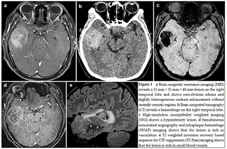
20170512093522 Figure 1 a Brain magnetic resonance imaging (MRI) reveals a 52 mm × 51 mm × 48 mm-lesion on the right temporal lobe and shows non-obvious edema and slightly heterogeneous contrast enhancement without centrally necrotic regions.b Brain computed tomography (CT) reveals a hemorrhage on the right temporal lobe. c High-resolution susceptibility weighted imaging (SWI) shows a hypointensity lesion.d Simultaneous noncontrast angiography and intraplaque hemorrhage (SNAP) imaging shows that the lesion is rich in vasculature.e T2 weighted inversion recovery based sequence for CSF suppression (T2 Flair)-imaging shows that the lesion is rich in small blood vessels.
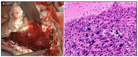
20170512093557 Figure 2 a The lesion,as observed during the surgical procedure,shows distinct boundaries with reddish brown appearance and is rich in venous vessels.b Histopathological analysis reveals that the tumor is composed of densely packed cells and shows nuclear and cell pleomorphism.The histopathological diagnosis is glioblastom.(Hematoxylin-eosin stain,×100)
[1]
Khanna A, Venteicher AS, Walcott BP, Kahle KT, Mordes DA, William CM, Ghogawala Z, Ogilvy CS. Glioblastoma mimicking an arteriovenous malformation. Front Neurol 2013, 4: 144.
[2]
Can SM, Aydin Y, Turkmenoglu O, Aydin F, Ziyal I. Giant cell glioblastoma manifesting as traumatic intracerebral hemorrhage—case report. Neurol Med Chir 2002, 42(12): 568-571.
[3]
Glass B, Abbott KH. Subarachnoid hemorrhage consequent to intracranial tumors: review of literature and report of seven cases. AMA Arch Neurol Psychiatry 1955, 73(4): 369-379.
[4]
Liwnicz BH, Wu SZ, Tew JM. The relationship between the capillary structure and hemorrhage in gliomas. J Neurosurg 1987, 66(4): 536-541.
[5]
Jain RK. Normalization of tumor vasculature: an emerging concept in antiangiogenic therapy. Science 2005, 307(5706): 58-62.
[6]
Takeshima H, Nishi T, Kuratsu J, Kamikubo Y, Kochi M, Ushio Y. Suppression of the tissue factor-dependent coagulation cascade: a contributing factor for the development of intratumoral hemorrhage in glioblastoma. Int J Mol Med 2000, 6(3): 271-276.
[7]
Schrader B, Barth H, Lang EW, Buhl R, Hugo HH, Biederer J, Mehdorn HM. Spontaneous intracranial haematomas caused by neoplasms. Acta Neurochir (Wien) 2000, 142(9): 979-985.
[8]
Maiuri F, D'Andrea F, Gallicchio B, Carandente M. Intracranial hemorrhages in metastatic brain tumors. J Neurosurg Sci 1985, 29(1): 37-41.
[9]
Schreuder T, Te Lintelo M, Kubat B, Koehler P. Anaplastic oligo-astrocytoma occurring after resection of a cerebral cavernous malformation; malignant transformation? Case report and review on etiology. J Neurol 2010, 257(3): 349-353.
[10]
Cemil B, Tun K, Polat O, Ozen O, Kaptanoglu E. Glioblastoma multiforme mimicking arteriovenous malformation. Turk Neurosurg 2009, 19(4): 433-436.
[11]
Jadik S, Stan AC, Dietrich U, Pietilä TA, Elsharkawy AE. Intraparenchymal meningioma mimicking cavernous malformation: a case report and review of the literature. J Med Case Rep 2014, 8: 467.
[12]
Tumialán LM, Brat DJ, Fountain AJ, Barrow DL. An astroblastoma mimicking a cavernous malformation: Case report. Neurosurgery 2007, 60(3): E569-E570.
[13]
Feiz-Erfan I, Zabramski JM, Herrmann LL, Coons SW. Cavernous malformation within a schwannoma: review of the literature and hypothesis of a common genetic etiology. Acta Neurochir (Wien) 2006, 148(6): 647-652.
[14]
Furuse M, Miyatake SI, Kuroiwa T. Cavernous malformation after radiation therapy for astrocytoma in adult patients: report of 2 cases. Acta Neurochir (Wien) 2005, 147(10): 1097-1101.
[15]
Zhang JY, Ming ZY, Wu AH. Is cerebral cavernous malformation a pre-glioma lesion. Chin Med J 2012, 125(24): 4511-4513.
Cruz AS, Jeyamohan S, Moisi M, Tubbs RS, Page J, Chamiraju P, Tkachenko L, Rostad S, Newell DW. Duralbased cavernoma of the posterior cranial fossa mimicking a meningioma: A case report. Cureus 2016, 8(4): e560.
[18]
Yang IY, Yum MS, Kim EH, Choi HW, Yoo HW, Ko TS. Two cases of familial cerebral cavernous malformation caused by mutations in the CCM1 gene. Korean J Pediatr 2016, 59(6): 280-284.
[19]
Hanjani SA. The genetics of cerebrovascular malformations. J Stroke Cerebrovasc Dis 2002, 11(5): 279-287.
[1]
Zhenxing Sun, Dan Yuan, Yaxing Sun, Zhanquan Zhang, James Wang, Yi Guo, Guoqin Wang, Dongkang Liu, Peng Chen, Linkai Jing, Feng Yang, Peihai Zhang, Huifang Zhang, Youtu Wu, Wei Shi, Guihuai Wang. Fluorescein sodium use during spinal ependymoma resection[J]. 临床转化神经科学, 2017, 3(3): 123-134.
 2017, Vol. 3