摘要 Objective: We aimed to provide an alternative cell source for cell therapy in leukoaraiosis (LA). Methods: Olfactory ensheathing cells (OECs) from the olfactory bulb were isolated,cultured,and purified.Next,the lentivirus carrying human VEGF165 gene was constructed and transfected into OECs. Results: The proliferative capacity of primary OECs was strong.OECs were infected with different multiplicity of infection.The expression level of VEGF was confirmed by real-time PCR with specific primers for GAPDH and VEGF,indicating that the genetically engineered OECs-VEGF produced VEGF with functional activity. Conclusions: Our data showed that these engineered OECs-VEGF highly express functional VEGF and retain the characteristics of astrocytes and Schwann cells,providing an alternative cell source for cell therapy in LA.
Abstract: Objective: We aimed to provide an alternative cell source for cell therapy in leukoaraiosis (LA). Methods: Olfactory ensheathing cells (OECs) from the olfactory bulb were isolated,cultured,and purified.Next,the lentivirus carrying human VEGF165 gene was constructed and transfected into OECs. Results: The proliferative capacity of primary OECs was strong.OECs were infected with different multiplicity of infection.The expression level of VEGF was confirmed by real-time PCR with specific primers for GAPDH and VEGF,indicating that the genetically engineered OECs-VEGF produced VEGF with functional activity. Conclusions: Our data showed that these engineered OECs-VEGF highly express functional VEGF and retain the characteristics of astrocytes and Schwann cells,providing an alternative cell source for cell therapy in LA.
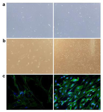
20170712150516 Figure 1 Morphology and identification of OECs isolated from rat olfactory bulbs. a. The primary OECs showed a typical spindlelike morphology with two or three processes or appearance (left picture: 40× magnification and right picture: 100× magnification). b. OECs formed colonies in the DF12 culture medium supplemented with 15% FBS, 2 μM forskolin, and 10 ng/mL bFGF (left picture: 40× magnification and right picture: 100× magnification). c. OECs were subjected for the immunofluorescence staining of p75 (Green) with Hoechst for nuclei labeling (left picture with 40× magnification and right picture with 100× magnification).
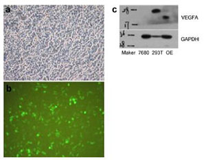
20170712150533 Figure 2 Construction of VEGF lentivirus. a–b. Human VEGF gene was cloned into the lentivirus vector with the marker of EGFP. The representative picture of transfected 293T cells with the VEGF lentivirus vector. c. The total protein of transfected 293T cells was subjected to western blotting for the VEGF specific antibody detection.
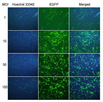
20170712150542 Figure 3 The infection efficiency with serial MOIs of control lentivirus. The OECs were infected with serial MOIs of control lentivirus: 1, 10, 50, and 100. The infection efficiency was monitored through fluorescence microscope with Hoechst for nuclei labeling.
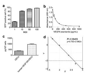
20170712150607 Figure 4 Quantification of infection efficiency for control lentivirus and the detection of secreted VEGF by OECs-VEGF. a. The percentage of GFP+ cells with different MOIs of control lentivirus: 1, 10, 50, and 100. b. The VEGF standards and their absorbance at 450 nm were plotted. c. Quantification of secreted VEGF by control OECs and OECs-VEGF. D. The log/log curve for standards with R2 = 0.99.
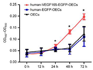
20170712150627 Figure 5 The functional activity of secreted VEGF by OECs-VEGF. The supernatant of cultivated OECs-VEGF was collected and the HUVECs were treated with the supernatant for up to 72 h. The OD450-OD460 values of HUVECs were compared between OECs, EGFP-OECs, and VEGF-OECs groups (* demonstrates P < 0.05).
[1]
Lin TC, Lin YH, Kao LL, Kao YH, Yang YH, Chou PS, Wu MN. An association between the location of white matter changes and the behavioral and psychological symptoms of dementia in Alzheimer's disease patients. Transl Neurosci Clin 2016, 2(1):8-16.
[2]
Sarabia-Cobo CM, Pérez V, Hermosilla C, Nuñez MJ, de Lorena P. Apathy and leukoaraiosis in mild cognitive impairment and Alzheimer's disease:Multicenter diagnostic criteria according to the latest studies. Dement Geriatr Cogn Dis Extra 2014, 4(2):228-235.
[3]
Schmidt R, Ropele S, Ferro J, Madureira S, Verdelho A, Petrovic K, Gouw A, van der Flier WM, Enzinger C, Pantoni L, et al. Diffusion-weighted imaging and cognition in the leukoariosis and disability in the elderly study. Stroke 2010, 41(5):e402-e408.
[4]
Brown WR, Thore CR. Review:Cerebral microvascular pathology in ageing and neurodegeneration. Neuropathol Appl Neurobiol 2011, 37(1):56-74.
[5]
Dehghanian F, Hojati Z, Kay M. New insights into VEGF-A alternative splicing:Key regulatory switching in the pathological process. Avicenna J Med Biotechnol 2014, 6(4):192-199.
[6]
Liu F, Ni JJ, Huang JJ, Kou ZW, Sun FY. VEGF over enhances the accumulation of phospho-S292 MeCP2 in reactive astrocytes in the adult rat striatum following cerebral ischemia. Brain Res 2015, 1599:32-43.
[7]
Vincent AJ, Taylor JM, Choi-Lundberg DL, West AK, Chuah MI. Genetic profile of olfactory ensheathing cells is distinct from that of Schwann cells and astrocytes. Glia 2005, 51(2):132-147.
[8]
Ramón-Cueto A. Olfactory ensheathing glia for nervous system repair. Exp Neurol 2011, 229(1):1.
[9]
Wang DJ. Why does a little mean a lot when you have nothing? A brief review of cell therapy strategies for spinal cord injury. Transl Neurosci Clin 2015, 1(2):102-109.
[10]
Cao L, Liu L, Chen ZY, Wang LM, Ye JL, Qiu HY, Lu CL, He C. Olfactory ensheathing cells genetically modified to secrete GDNF to promote spinal cord repair. Brain 2004, 127(3):535-549.
[11]
Wewetzer K, Verdú E, Angelov DN, Navarro X. Olfactory ensheathing glia and Schwann cells:Two of a kind? Cell Tissue Res 2002, 309(3):337-345.
[12]
Ali N, Yoshizumi M, Fujita Y, Izawa Y, Kanematsu Y, Ishizawa K, Tsuchiya K, Yano S, Sone S, Tamaki T. A novel Src kinase inhibitor, M475271, inhibits VEGF-induced human umbilical vein endothelial cell proliferation and migration. J Pharmacol Sci 2005, 98(2):130-141.
[13]
Rousseau S, Houle F, Landry J, Huot J. p38 MAP kinase activation by vascular endothelial growth factor mediates actin reorganization and cell migration in human endothelial cells. Oncogene 1997, 15(18):2169-2177.
[14]
McMullen M, Keller R, Sussman M, Pumiglia K. Vascular endothelial growth factor-mediated activation of p38 is dependent upon Src and RAFTK/Pyk2. Oncogene 2004, 23(6):1275-1282.
[15]
Morimoto T, Yasuhara T, Kameda M, Baba T, Kuramoto S, Kondo A, Takahashi K, Tajiri N, Wang FF, Meng J, et al. Striatal stimulation nurtures endogenous neurogenesis and angiogenesis in chronic-phase ischemic stroke rats. Cell Transplant 2011, 20(7):1049-1064.
[16]
Adib-Samii P, Devan W, Traylor M, Lanfranconi S, Zhang CR, Cloonan L, Falcone GJ, Radmanesh F, Fitzpatrick K, Kanakis A, et al. Genetic architecture of white matter hyperintensities differs in hypertensive and nonhypertensive ischemic stroke. Stroke 2015, 46(2):348-353.
[17]
Crafts TD, Jensen AR, Blocher-Smith EC, Markel TA. Vascular endothelial growth factor:Therapeutic possibilities and challenges for the treatment of ischemia. Cytokine 2015, 71(2):385-393.
[18]
Bouleti C, Mathivet T, Coqueran B, Serfaty JM, Lesage M, Berland E, Ardidie-Robouant C, Kauffenstein G, Henrion D, Lapergue B, et al. Protective effects of angiopoietin-like 4 on cerebrovascular and functional damages in ischaemic stroke. Eur Heart J 2013, 34(47):3657-3668.
[19]
Grueter BE, Schulz UG. Age-related cerebral white matter disease (leukoaraiosis):A review. Postgrad Med J 2012, 88(1036):79-87.
[20]
Ekberg JAK, St John JA. Crucial roles for olfactory ensheathing cells and olfactory mucosal cells in the repair of damaged neural tracts. Anat Rec 2014, 297(1):121-128.
[21]
Bartolomei JC, Greer CA. Olfactory ensheathing cells:Bridging the gap in spinal cord injury. Neurosurgery 2000, 47(5):1057-1069.
[22]
Mouhieddine TH, Kobeissy FH, Itani M, Nokkari A, Wang KKW. Stem cells in neuroinjury and neurodegenerative disorders:Challenges and future neurotherapeutic prospects. Neural Regen Res 2014, 9(9):901-906.
 2017, Vol. 3