Clinical features and prognostic factors of primary intracranial malignant fibrous histiocytoma: A report of 8 cases and a literature review
Peng Li1, Qiangyi Zhou1, Zhijun Yang1, Zhenmin Wang1, Shiwei Li1, Xingchao Wang1, Bo Wang1, Fu Zhao2, Pinan Liu1,2
1 Department of Neurosurgery, Beijing Tiantan Hospital, Capital Medical University, Beijing 100050, China;
2 Department of Neural Reconstruction, Beijing Neurosurgery Institute, Capital Medical University, Beijing 100050, China
摘要 Objective: Primary intracranial malignant fibrous histiocytoma (MFH) is rare. We describe the detailed clinical features of 8 cases and fully review the literature to evaluate several prognostic factors. Methods: Eight patients with pathologically confirmed primary intracranial MFH were retrospectively reviewed. We searched PubMed for relevant articles with the term "intracranial malignant fibrous histiocytoma". Results: Of the 8 patients, 4 were men and 4 were women. Three patients had received previous radiotherapy. The age of the patients ranged from 19 to 69 years, with a median age of 48 years. Most tumors could be totally resected; and only 1 tumor was subtotally resected. Six patients received postoperative radiotherapy and 3 patients received postoperative chemotherapy. Most patients died within the first year after surgery; and only 1 patient was still alive on the date of the last follow-up. We reviewed the literature and included a total of 46 patients in the Kaplan-Meier survival analysis. Young patients (less than 30 years old) seemed to have a better prognosis and survival rate than older patients (more than 30 years old) (log-rank test, P=0.008). However, sex (P=0.675), extent of resection (P=0.934), postoperative radiotherapy (P=0.592), and postoperative chemotherapy (P=0.424) did not affect patient prognosis. Conclusions: The prognosis of MFH is usually poor, and most patients die within the first year after surgery. Younger MFH patients (less than 30 years old) seem to have a better prognosis and improved survival compared to older patients.
Abstract: Objective: Primary intracranial malignant fibrous histiocytoma (MFH) is rare. We describe the detailed clinical features of 8 cases and fully review the literature to evaluate several prognostic factors. Methods: Eight patients with pathologically confirmed primary intracranial MFH were retrospectively reviewed. We searched PubMed for relevant articles with the term "intracranial malignant fibrous histiocytoma". Results: Of the 8 patients, 4 were men and 4 were women. Three patients had received previous radiotherapy. The age of the patients ranged from 19 to 69 years, with a median age of 48 years. Most tumors could be totally resected; and only 1 tumor was subtotally resected. Six patients received postoperative radiotherapy and 3 patients received postoperative chemotherapy. Most patients died within the first year after surgery; and only 1 patient was still alive on the date of the last follow-up. We reviewed the literature and included a total of 46 patients in the Kaplan-Meier survival analysis. Young patients (less than 30 years old) seemed to have a better prognosis and survival rate than older patients (more than 30 years old) (log-rank test, P=0.008). However, sex (P=0.675), extent of resection (P=0.934), postoperative radiotherapy (P=0.592), and postoperative chemotherapy (P=0.424) did not affect patient prognosis. Conclusions: The prognosis of MFH is usually poor, and most patients die within the first year after surgery. Younger MFH patients (less than 30 years old) seem to have a better prognosis and improved survival compared to older patients.
Peng Li, Qiangyi Zhou, Zhijun Yang, Zhenmin Wang, Shiwei Li, Xingchao Wang, Bo Wang, Fu Zhao, Pinan Liu. Clinical features and prognostic factors of primary intracranial malignant fibrous histiocytoma: A report of 8 cases and a literature review[J]. 临床转化神经科学, 2016, 2(3): 155-164.
Peng Li, Qiangyi Zhou, Zhijun Yang, Zhenmin Wang, Shiwei Li, Xingchao Wang, Bo Wang, Fu Zhao, Pinan Liu. Clinical features and prognostic factors of primary intracranial malignant fibrous histiocytoma: A report of 8 cases and a literature review. Translational Neuroscience and Clinics, 2016, 2(3): 155-164.
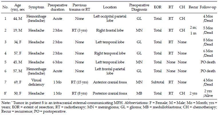
20161111172835 Table 1 Clinical features of 8 cases with primary intracranial malignant fibrous histiocytoma
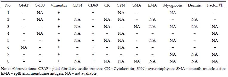
20161111172848 Table 2 Results of immunohistochemical staining in 8 cases
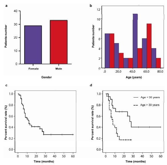
20161111172903 Figure 1 (a, b) Bar graph depicting the sex and age distribution in a total of 62 MFH patients. (c) Kaplan-Meier plots of overall survival for 46 MFH patients. (d) Kaplan-Meier plots of overall survival for MFH patients older (dotted line) or younger (solid line) than the age of 30 years old (log-rank test, P= 0.008).
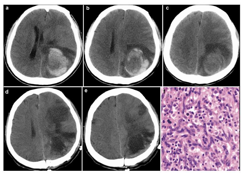
20161116191142 Figure 2 Case 1. Preoperative cranial CT shows an evident hematoma in the left occipital parietal lobe (a–c). Postoperative cranial CT shows total resection of the tumor (d & e). Pathological examination (f) reveals the diagnosis of MFH. (Hematoxylin & eosin staining, ×100)
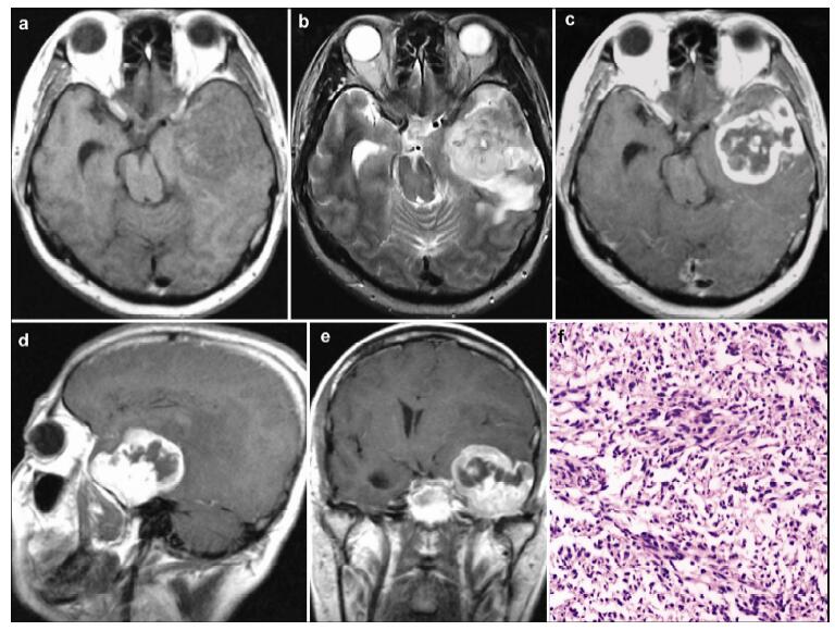
20161116191301 Figure 3 Case 5. Preoperative cranial magnetic resonance imaging (MRI) shows a tumor lesion in the left temporal lobe. The tumor is isointense on T1-weighted image (a), and hyperintense on T2-weighted image (b). A peritumoral edema can be observed, and the brain stem is compressed. The tumor is heterogeneously enhanced after contrast administration (c–e). Pathological examination (f) reveals the diagnosis of MFH. (Hematoxylin & eosin staining, ×100)
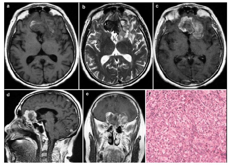
20161116191408 Figure 4 Case 7. Preoperative cranial magnetic resonance imaging (MRI) shows a tumor lesion located at the anterior cranial fossa. The tumor is isointense on T1-weighted image (a), and hypointense on T2-weighted image (b). The tumor is heterogeneously enhanced after contrast administration (c–e). Pathological examination (f) reveals the diagnosis of MFH. (Hematoxylin & eosin staining, ×100)
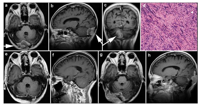
20161116191520 Figure 5 Case 8. Preoperative cranial magnetic resonance imaging (MRI) shows a tumor lesion (arrow) in the posterior cranial fossa extending outward to the subcutaneous tissues (a-c). Pathological examination (d) of the resected tumor reveals the diagnosis of MFH. (Hematoxylin & eosin staining, ×100) Postoperative cranial MRI shows no remnant of the tumor (e, f). Two years after the operation, cranial MRI shows recurrence of the tumor, mainly in the subcutaneous tissues (g, h).
[1]
Pimentel J, Fernandes A, Távora L, Miguéns J, Lobo Antunes J. Benign isolated fibrohistiocytic tumor arising from the central nervous system. Considerations about two cases. Clin Neuropathol 2002, 21(3):93-98.<br />
[2]
Deb P, Singh V, Dutta V, Bhatoe HS, Chandran VM. Primary intracranial benign fibrous histiocytoma:Report of an unusual case. J Cancer Res Ther 2014, 10(1):200-202.<br />
[3]
Moliterno JA, Sood S, Zambrano E, Kim JH, Piepmeier JM, Baehring JM. Intracranial benign fibrous histiocytomas:A case report and review. J Neurooncol 2009, 92(2):203-209.<br />
[4]
Hayami J, Kurokawa I, Hashimoto K, Kusumoto K. Malignant fibrous histiocytoma of scalp with intracranial invasion 20 years after postoperative irradiation. J Craniofac Surg 2003, 14(1):74-77.<br />
[5]
Hamlat A, Adn M, Caulet-Maugendre S, Guegan Y. Cerebellar malignant fibrous histiocytoma:Case report and literature review. Neurosurgery 2004, 54(3):745-751.<br />
[6]
Gelabert-González M, Fernández-Villa JM, Reyes-Santías R. Histiocitoma fibroso maligno de duramadre. Neurocirugía 2003, 14(3):235-239.<br />
[7]
Broniscer A, Ke W, Fuller CE, Wu J, Gajjar A, Kun LE. Second neoplasms in pediatric patients with primary central nervous system tumors:The St. Jude Children's Research Hospital experience. Cancer 2004, 100(10):2246-2252.<br />
[8]
Mitsuhashi T, Watanabe M, Ohara Y, Hatashita S, Ueno H. Multifocal primary intracerebral malignant fibrous histiocytoma-Case report. Neurol Med Chir (Tokyo) 2004, 44(5):249-254.<br />
[9]
Maruno M, Ghulam Muhammad AKM, Taguchi J, Suzuki T, Wada K, Isaka T, Yoshimine T. Giant cell type of primary intracranial malignant fibrous histiocytoma:A case report. Brain Tumor Pathol 2006, 23(1):65-70.<br />
[10]
Harries AM, Mitchell R. Haemorrhagic cerebellar fibrous histiocytoma:Case report and literature review. Br J Neurosurg 2011, 25(1):120-121.<br />
[11]
Sarrami AH, Setareh M, Afshar-Moghaddam N, Izadinejad M, Saadatnia M. A case of intracranial malignant fibrous histiocytoma. J Res Med Sci 2011, 16(7):968-973.<br />
[12]
Gelincik I. Cerebellar malignant fibrous histiocytoma. Indian J Pathol Microbiol 2012, 55(3):402-405.<br />
[13]
Ozdemir M, Ozgural O, Bozkurt M, Torun FM, Heper AO, Tuna H. Primary intracerebral malignant fibrous histiocytoma mimicking a meningioma. Turk Neurosurg 2012, 22(4):475-477.<br />
[14]
Grahovac G, Chudy D, Heinrich Z, Zarkovic K. Implantation metastasis of malignant fibrous histiocytoma along the stereotactic biopsy tract. Clin Neurol Neurosurg 2013, 115(7):1160-1161.<br />
[15]
Yoo RE, Choi SH, Park SH, Jung HW, Kim JH, Sohn CH, Chang KH. Primary intracerebral malignant fibrous histio-cytoma:CT, MRI, and PET-CT findings. J Neuroimaging 2013, 23(1):141-144.<br />
[16]
Kurosaki M, Kambe A, Ishibashi M, Watanabe T, Horie Y. A case report of sarcoma of the sella caused by postoperative radiotherapy for a prolactin-producing pituitary adenoma. Brain Tumor Pathol 2014, 31(3):187-191.<br />
[17]
Özhan S, Tali ET, I?ik S, Saygili MR, Baykaner K. Haematoma-like primary intracranial malignant fibrous histiocytoma in a 5-year-old girl. Neuroradiology 1999, 41(7):523-525.<br />
[18]
Fritz MA, Sade B, Bauer TW, Wood BG, Lee JH. Benign fibrous histiocytoma of the pterygopalatine fossa with intracranial extension. Acta Neurochir 2006, 148(1):73-76.<br />
[19]
Graber JJ, Nayar A, Zagzag D. Metastatic cerebral malignant fibrous histiocytoma masquerading as neurocysticercosis. J Neurooncol 2011, 105(2):437-439.<br />
[20]
Schrader B, Holland BR, Friedrichsen C. Rare case of a primary malignant fibrous histiocytoma of the brain. Neuroradiology 1989, 31(2):177-179.<br />
[21]
Mahore A, Ramdasi R, Dange N, Epari S. Malignant fibrous histiocytoma of the skull base:A neurosurgical nuance. Asian J Neurosurg 2015, 10(2):135-138.<br />
[22]
Wang J, Zhong WM, Xu YH, Feng L, Li Y, Dong B. A primary malignant fibrous histiocytoma of the scalp and intracranial tumor bleeding:A case report. J Med Case Rep 2014, 8:50.<br />
[23]
Matsuura S, Takagi T, Tan EC, Mizuno S, Imagunbai N, Hasegawa R. Malignant fibrous histiocytoma of the occipital bone with intracranial extension-Case report. Neurol Med Chir (Tokyo) 1991, 31(4):219-222.<br />
[24]
Wu TH, Shih CW, Huang JS, Wang CH, Yeh KY. Unusual hematogenous brain metastasis in malignant fibrous histio-cytoma of the maxillary sinus. Int J Clin Oncol 2012, 17(1):69-74.<br />
[25]
Itoyama Y, Nagahiro S, Seto H, Sueyoshi N, Kuratsu JI, Ushio Y. Brain metastasis from malignant fibrous histiocytoma of the heart:Case report. Neurosurgery 1990, 26(4):692-695.<br />
[26]
Gonzalez-Vitale JC, Slavin RE, McQueen JD. Radiation-induced intracranial malignant fibrous histiocytoma. Cancer 1976, 37(6):2960-2963.<br />
[27]
Baehring JM, Alemohammed S, Croul SE. Malignant fibrous histiocytoma presenting as an intraventricular mass five years after incidental detection of a mass lesion. J Neurooncol 2001, 52(2):157-160.<br />
[28]
Hirato J, Nakazato Y, Sasaki A, Yokota M, Nojiri K, Toyoda O, Nakajima H. Intracranial malignant fibrous histiocytoma:Characterization of GFAP-positive cells in the tumor. Clin Neuropathol 1994, 13(6):315-322.<br />
[29]
Amendola BE, Amendola MA, McClatchey KD. Radiation-induced malignant fibrous histiocytoma:A report of five cases including two occurring post whole brain irradiation. Cancer Invest 1985, 3(6):507-513.<br />
[30]
Martinez-Salazar A, Supler M, Rojiani AM. Primary intracerebral malignant fibrous histiocytoma:Immunohisto-chemical findings and etiopathogenetic considerations. Mod Pathol 1997, 10(2):149-154.<br />
[31]
Paulus W, Peiffer J, Grote E. Intracerebral malignant fibrous histiocytoma at site of a previously excised low grade glioma. Acta Neurochir (Wien) 1989, 99(3-4):161-165.<br />
[32]
Bora H, Oztürk B, Akmansu M, Yenidunya S, Egehan I. Intracerebral malignant fibrous histiocytoma in a 5-year-old girl. Radiat Med 1999, 17(5):355-358.<br />
[33]
Kalyanaraman UP, Taraska JJ, Fierer JA, Elwood PW. Malignant fibrous histiocytoma of the meninges. Histological, ultrastructural, and immunocytochemical studies. J Neurosurg 1981, 55(6):957-962.<br />
[34]
Akimoto J, Takeda Y, Hasue M, Ito H, Kiguchi E. Primary meningeal malignant fibrous histiocytoma with cerebrospinal dissemination and pulmonary metastasis. Acta Neurochir (Wien) 1998, 140(11):1191-1196.<br />
[35]
Ho YS, Wei CH, Tsai MD, Wai YY. Intracerebral malignant fibrous histiocytoma:Case report and review of the literature. Neurosurgery 1992, 31(3):567-570.<br />
[36]
Berry AD 3rd, Reintjes SL, Kepes JJ. Intracranial malignant fibrous histiocytoma with abscess-like tumor necrosis. Case report. J Neurosurg 1988, 69(5):780-784.<br />
[37]
Kepes JJ. "Xanthomathous" lesions of the central nervous system:Definition, classification and some recent observations. In Progress in Neuropathology Vol. 4. Zimmerman HMN, Ed. New York:Raven, 1979, pp 179-213.<br />
[38]
Akai T, Yamamoto K, Iida T, Iizuka H, Nojima T. Malignant fibrous histiocytoma in the craniocervical junction presenting with severe occipitalgia. Brain Tumor Pathol 2006, 23(2):101-105.<br />
[39]
Le Doussal V, Coindre JM, Leroux A, Hacene K, Terrier P, Bui NB, Bonichon F, Collin F, Mandard AM, Contesso G. Prognostic factors for patients with localized primary malignant fibrous histiocytoma:A multicenter study of 216 patients with multivariate analysis. Cancer 1996, 77(9):1823-1830.<br />
[40]
Fujimura N, Sugita Y, Hirohata M, Naohisa M, Terasaki M, Tokutmi T, Shigemori M. Primary intracerebral malignant fibrous histiocytoma in a child. Pediatr Neurosurg 2002, 37(5):271-274.<br />
[41]
Pople IK, Harding B. Primary intracranial malignant fibrous histiocytoma in a 5-year-old boy:Case report. Br J Neurosurg 1991, 5(5):509-513.<br />
 2016, Vol. 2