Surgical resection of a cervical intramedullary schwannoma:A case report and literature review
Peihai Zhang1, Zhenxing Sun1, Dan Yuan2, Yaxing Sun3, Zhanquan Zhang4, James Wang1, Yi Guo1, Guoqin Wang1, Dongkang Liu1, Peng Chen1, Linkai Jing1, Feng Yang1, Huifang Zhang1, Wei Shi1, Guihuai Wang1
1 Department of Neurosurgery, Changgung Hospital, Medical Center, Tsinghua University, Beijing 102218, China;
2 Department of Nephrology, Beijing Luhe Hospital, Capital Medical University, Beijing 101149, China;
3 Department of Psychiatry, Zaozhuang Mental Health Center, Zaozhuang 277103, China;
4 Department of Neurosurgery, The Fifth People's Hospital of Datong, Regional Medical Center of Shanxi Province, Datong 037006, China
摘要 Schwannomas are the most common type of spinal tumor, and they most commonly occur in intradural extramedullary locations. Intramedullary schwannomas of the central nervous system are very rare and are difficult to diagnose using preoperative imaging. Here, we report a rare, tiny cervical intramedullary schwannoma and review the literature regarding the clinical presentation, magnetic resonance imaging, pathology, and surgical experience associated with this rare tumor type.
Abstract: Schwannomas are the most common type of spinal tumor, and they most commonly occur in intradural extramedullary locations. Intramedullary schwannomas of the central nervous system are very rare and are difficult to diagnose using preoperative imaging. Here, we report a rare, tiny cervical intramedullary schwannoma and review the literature regarding the clinical presentation, magnetic resonance imaging, pathology, and surgical experience associated with this rare tumor type.
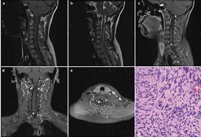
20171023160911 Figure 1 (a) Sagittal T1-weighted magnetic resonance image demonstrating an isointense lesion. (b) Sagittal T2-weighted magnetic resonance image demonstrating a slightly hyperintense lesion, with swelling surrounding it. (c–e) Sagittal, coronal, and axial T1-weighted magnetic resonance images, with gadolinium contrast, demonstrating homogeneous enhancement of the tumor. The tumor appears as a solid mass, located to the right side of spinal cord, with clear margins. (f) Hematoxylin and eosin staining from the biopsy of the mass showing bipolar spindle cells with nuclei arranged in a palisade pattern (Hematoxylin & Eosin stain; magnification, ×100).
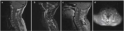
20171023160923 Figure 2 Post-operative magnetic resonance image; no recurrence of the solid tumor is evident.
[1]
Levy WJ, Latchaw J, Hahn JF, SawhnyB, Bay J, Dohn DF. Spinal neurofibromas:a report of 66 cases and a comparison with meningiomas. Neurosurgery 1986, 18(3):331-334.
[2]
Ross DA, Edwards MSB, Wilson CB. Intramedullary neurilemomas of the spinal cord:report of two cases and review ofthe literature. Neurosurgery 1986, 19(3):458-464.
[3]
Ho T, Tai KS, Fan YW, Leong LL. Intramedullary spinal schwannoma:case report and review of preoperative magnetic resonance imaging features. Asia J Surg 2006, 29(4):306-308.
[4]
Penfield W. Cytology and Cellular Pathology of the Nervous System. New York:Paul B Hoeber Inc., 1932.
[5]
Nicoletti GF, Passanisi M, Castana L, Albanese V. Intramedullary spinal neurinoma:case report and review of 46 cases. J Neurosurgery Sci 1994, 38(3):187-191.
[6]
Karatay M, Koktekir E, Erdem Y, Celik H, Sertbas I, Bayar MA. Intramedullary schwannoma of conus medullaris with syringomyelia.Asia J Surg2017, 40(3):240-242.
[7]
Sun B, Che XM, Gu SX, Liu XD, Shou JJ, Gu WT. The clinical diagnosis and treatment of intramedullary spinal schwannomas. Chin J Neurosurg 2012, 28(6):577-580. (in Chinese)
[8]
Colosimo C, Cerase A, Denaro L, Maira G, Greco R. Magnetic resonance imaging of intramedullary spinal cord schwannomas. J Neurosurg 2003, 99(1):114-117.
[9]
Kodama Y, Terae S, Hida K, Chu BC, Kaneko K, Miyasaka K. Intramedullary schwannoma of the spinal cord:report of two cases. Neuroradiology 2001, 43(7):567-571.
[10]
Conti P, Pansini G, Mouchaty H, Capuano C, Conti R. Spinal neurinomas:retrospective analysis and long-term outcome of 179 consecutively operated cases and review of the literature. Surg Neurol2004, 61(1):34-43.
[11]
Shenoy SN, Raja A. Cystic cervical intramedullary schwannoma with syringomyelia. Neurol India2005, 53(2):224-225.
[12]
Riffaud L, Morandi X, Massengo S, Carsin-Nicol B, Heresbach N, Guegan Y. MRI of intramedullary spinal schwannomas:case report and review of the literature. Neuroradiology2000, 42(4):275-279.
 2017, Vol. 3