Post-traumatic cerebrospinal fluid rhinorrhea associated with craniofacial fibrous dysplasia: Case report and literature review
Peng Li1, Qiangyi Zhou1, Zhijun Yang1, Zhenmin Wang1, Shiwei Li1, Xingchao Wang1, Bo Wang1, Fu Zhao2, Pinan Liu1,2
1 Department of Neurosurgery, Beijing Tiantan Hospital, Capital Medical University, Beijing 100050, China;
2 Department of Neural Reconstruction, Beijing Neurosurgery Institute, Capital Medical University, Beijing 100050, China
摘要 Objective: Fibrous dysplasia (FD) is an unusual developmental abnormality of the skeleton. When facial and cranial bones are involved in FD, it is termed craniofacial fibrous dysplasia (CFD). Although several reports have reported that CFD has a tendency for spontaneous cerebrospinal fluid (CSF) leakage, there have been no related English-language case reports. We present the first case of post-traumatic CSF rhinorrhea associated with CFD. Methods: A 30-year-old man presented with CSF rhinorrhea after a mild head trauma. Computed tomography cisternogram located a defect in the posterior wall of the right frontal sinus. Imaging examination also showed the evident expansion of multiple skull bones, spinal scoliosis, and multiple local enlargements of ribs. Without café-au-lait cutaneous spots and endocrine abnormalities, polyostotic FD was diagnosed instead of McCune-Albright syndrome (MAS). The patient underwent craniotomy fistula repair surgery. The excised bone was contoured to be thinner to increase the cranial cavity. The patient recovered well and CSF leakage did not recur. But during a nineteen-month follow up, sight in the patient's left eye was decreased. MAS was suspected. Unfortunately the patient refused to take the proposed decompression surgery and laboratory tests of serum hormones. Conclusions: CFD, if the wall of the paranasal sinus is involved and the cranial cavity is decreased, may increase the risk of CSF rhinorrhea after head trauma. Expectant management is recommended in asymptomatic CFD patients even in the presence of optic nerve compression. As MAS may cause more problems, it should be precluded before polyostotic FD is diagnosed.
Abstract: Objective: Fibrous dysplasia (FD) is an unusual developmental abnormality of the skeleton. When facial and cranial bones are involved in FD, it is termed craniofacial fibrous dysplasia (CFD). Although several reports have reported that CFD has a tendency for spontaneous cerebrospinal fluid (CSF) leakage, there have been no related English-language case reports. We present the first case of post-traumatic CSF rhinorrhea associated with CFD. Methods: A 30-year-old man presented with CSF rhinorrhea after a mild head trauma. Computed tomography cisternogram located a defect in the posterior wall of the right frontal sinus. Imaging examination also showed the evident expansion of multiple skull bones, spinal scoliosis, and multiple local enlargements of ribs. Without café-au-lait cutaneous spots and endocrine abnormalities, polyostotic FD was diagnosed instead of McCune-Albright syndrome (MAS). The patient underwent craniotomy fistula repair surgery. The excised bone was contoured to be thinner to increase the cranial cavity. The patient recovered well and CSF leakage did not recur. But during a nineteen-month follow up, sight in the patient's left eye was decreased. MAS was suspected. Unfortunately the patient refused to take the proposed decompression surgery and laboratory tests of serum hormones. Conclusions: CFD, if the wall of the paranasal sinus is involved and the cranial cavity is decreased, may increase the risk of CSF rhinorrhea after head trauma. Expectant management is recommended in asymptomatic CFD patients even in the presence of optic nerve compression. As MAS may cause more problems, it should be precluded before polyostotic FD is diagnosed.
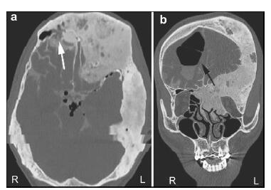
20161117200932 Figure 1 (a, b) A computed tomography (CT) scan demonstrating the expansion of multiple skull bones. The involved bones present with various appearances of mixed radio-dense/radio-lucent lesions, which can be described as “ground-glass”, “lytic”, and “cystic”. Left: Axial CT cisternography demonstrates a fistula at the posterior wall of the right frontal sinus (white arrow). The density of both frontal involved bones is slightly lower than for other skull bones. Right: A coronal CT scan demonstrates pneumocephalus in the right frontal lobe (black arrow).
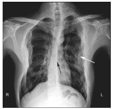
20161117200955 Figure 2 X-ray chest analysis demonstrating scoliosis (black arrow) and multiple local enlargements of the ribs (white arrow).
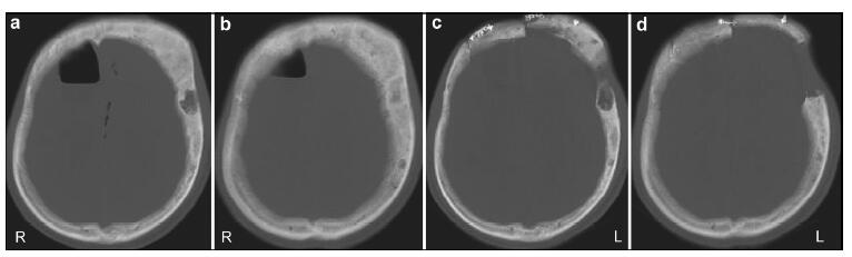
20161117201031 Figure 3 A CT scan demonstrating a contoured bone and enlarged cranial cavity. (a, b) Before the fistula repair surgery. (c, d) After the fistula repair surgery.
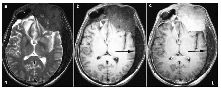
20161117201119 Figure 4 Axial MRI demonstrating abnormal thickening of the bones in a patient with FD. (a) T2-weighted images show the involved skull bones are hypointense. (b, c) T1-weighted images show the involved skull bones are hypointense (b) with varying enhancement (c). Left frontal bone and the wall of the right frontal sinus showed evident enhancement (white arrows) while the left posterior skull bones did not (black arrows).
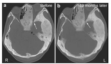
20161117201145 Figure 5 (a, b) CT showing the left optic canal before and nineteen months after surgery (arrows). The compression of the left optic nerve was more severe at nineteen months after surgery (right) than before surgery (left).
[1] Hanifi B, Samil KS, Yasar C, Cengiz C, Ercan A, Ramazan D. Craniofacial fibrous dysplasia. Clin Imaging 2013, 37(6):1109-1115.
[2] Chapurlat RD, Orcel P. Fibrous dysplasia of bone and McCune-Albright syndrome. Best Pract Res Clin Rheumatol 2008, 22(1):55-69.
[3] Jee WH, Choi KH, Choe BY, Park JM, Shinn KS. Fibrous dysplasia:MR imaging characteristics with radiopathologic-correlation. AJR Am J Roentgenol 1996, 167(6):1523-1527.
[4] Assaf AT, Benecke AW, Riecke B, Zustin J, Fuhrmann AW, Heiland M, Friedrich RE. Craniofacial fibrous dysplasia (CFD) of the maxilla in an 11-year old boy:A case report. J Craniomaxillofac Surg 2012, 40(8):788-792.
[5] Lee JS, FitzGibbon EJ, Chen YR, Kim HJ, Lustig LR, Akintoye SO, Collins MT, Kaban LB. Clinical guidelines for the management of craniofacial fibrous dysplasia. Orphanet J Rare Dis 2012, 7(Suppl 1):S2.
[6] Menon S, Venkatswamy S, Ramu V, Banu K, Ehtaih S, Kashyap VM. Craniofacial fibrous dysplasia:Surgery and literature review. Ann Maxillofac Surg 2013, 3(1):66-71.
[7] Valentini V, Cassoni A, Marianetti TM, Terenzi V, Fadda MT, Iannetti G. Craniomaxillofacial fibrous dysplasia:Conservative treatment or radical surgery? A retrospective study on 68 patients. Plast Reconstr Surg 2009, 123(2):653-660.
[8] Diah E, Morris DE, Lo LJ, Chen YR. Cyst degeneration in craniofacial fibrous dysplasia:Clinical presentation and management. J Neurosurg 2007, 107(3):504-508.
[9] Wei YT, Jiang S, Cen Y. Fibrous dysplasia of skull. J Craniofac Surg 2010, 21(2):538-542.
[10] Cai MJ, Ma LT, Xu GZ, Gruen P, Li J, Yang M, Pan L, Guan HF, Chen G, Gong J, et al. Clinical and radiological observation in a surgical series of 36 cases of fibrous dysplasia of the skull. Clin Neurol Neurosurg 2012, 114(3):254-259.
[11] Ganda K, Seibel MJ. Rapid biochemical response to denosumab in fibrous dysplasia of bone:Report of two cases. Osteoporos Int 2014, 25(2):777-782.
[12] Riddle ND, Bui MM. Fibrous dysplasia. Arch Pathol Lab Med 2013, 137(1):134-138.
[13] Pai B, Ferdinand D. Fibrous dysplasia causing safeguarding concerns. Arch Dis Child 2013, 98(12):1003.
[14] Kamochi H, Kusaka G, Ishikawa M, Ishikawa S, Tanaka Y. Late onset cerebrospinal fluid leakage associated with past head injury. Neurol Med Chir (Tokyo) 2013, 53(4):217-220.
[15] Ziu M, Savage JG, Jimenez DF. Diagnosis and treatment of cerebrospinal fluid rhinorrhea following accidental traumatic anterior skull base fractures. Neurosurg Focus 2012, 32(6):E3.
[16] Kong F, Zhang QH, Qu QY. Transclival cerebrospinal fluid rhinorrhea as the initial presenting symptom of a tiny intraduralchordoma. J Clin Neurosci 2010, 17(8):1083-1085.
[17] Hsu CCT, Kwan GNC, Bhuta SS. Non-traumatic cerebro-spinal fluid rhinorrhea caused by ethmoid sinus osteoma. J Clin Neurosci 2010, 17(9):1185-1186.
[18] Lam G, Mehta V, Zada G. Spontaneous and medically induced cerebrospinal fluid leakage in the setting of pituitary adenomas:Review of the literature. Neurosurg Focus 2012, 32(6):E2.
[19] Perani D, Scotti G, Colombo N, Sterzi R, Castelli A. Spontaneous CSF rhinorrhea through the lamina cribrosa associated with primary empty sella. Ital J Neurol Sci 1984, 5(2):167-172.
[20] Wang EW, Vandergrift WA, Schlosser RJ. Spontaneous CSF Leaks. Otolaryngol Clin North Am 2011, 44(4):845-856.
[21] Yang ZJ, Wang B, Wang CC, Liu P. Primary spontaneous cerebrospinal fluid rhinorrhea:A symptom of idiopathic intracranial hypertension? J Neurosurg 2011, 115(1):165-170.
[22] Schreiber A, Villaret AB, Maroldi R, Nicolai P. Fibrous dysplasia of the sinonasal tract and adjacent skull base. Curr Opin Otolaryngol Head Neck Surg 2012, 20(1):45-52.
[23] Feingold RS, Argamaso RV, Strauch B. Free fibula flap mandible reconstruction for oral obstruction secondary to giant fibrous dysplasia. Plast Reconstr Surg 1996, 97(1):196-201.
[24] Amit M, Collins MT, FitzGibbon EJ, Butman JA, Fliss DM, Gil Z. Surgery versus watchful waiting in patients with craniofacial fibrous dysplasia-A meta-analysis. PLoS One 2011, 6(9):e25179.
[25] Hansen MR, Moffat JC. Osteosarcoma of the skull base after radiation therapy in a patient with McCune-Albright syndrome:Case report. Skull Base 2003, 13(2):79-83.
[26] Cutler CM, Lee JS, Butman JA, FitzGibbon EJ, Kelly MH, Brillante BA, Feuillan P, Robey PG, DuFresne CR, Collins MT. Long-term outcome of optic nerve encasement and optic nerve decompression in patients with fibrous dysplasia:Risk factors for blindness and safety of observation. Neurosurgery 2006, 59(5):1011-1017.
[27] Lala R, Matarazzo P, Andreo M, Defilippi C, de Sanctis C. Impact of endocrine hyperfunction and phosphate wasting on bone in McCune-Albright syndrome. J Pediatr Endocrinol Metab 2002, 15 Suppl 3:913-920.
[28] Kollerova J, Koller T, Zelinkova Z, Kostalova L, Payer J. Treatment of pathological bone fractures in a patient with McCune-Albright syndrome. Case Rep Endocrinol 2013, 2013:589872.
 2016, Vol. 2