| Original Articles |
|


|
|
|
| Clinical features and prognostic factors of primary intracranial malignant fibrous histiocytoma: A report of 8 cases and a literature review |
| Peng Li1, Qiangyi Zhou1, Zhijun Yang1, Zhenmin Wang1, Shiwei Li1, Xingchao Wang1, Bo Wang1, Fu Zhao2, Pinan Liu1,2 |
1 Department of Neurosurgery, Beijing Tiantan Hospital, Capital Medical University, Beijing 100050, China;
2 Department of Neural Reconstruction, Beijing Neurosurgery Institute, Capital Medical University, Beijing 100050, China |
|
|
|
|
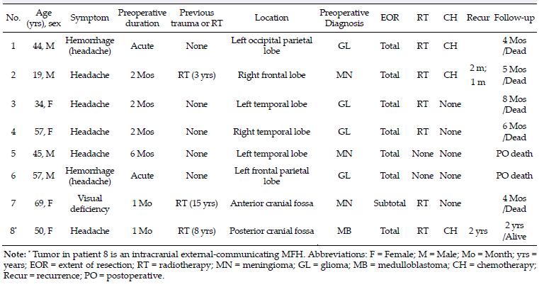
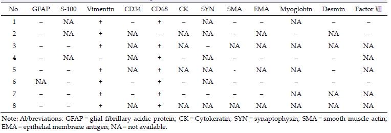
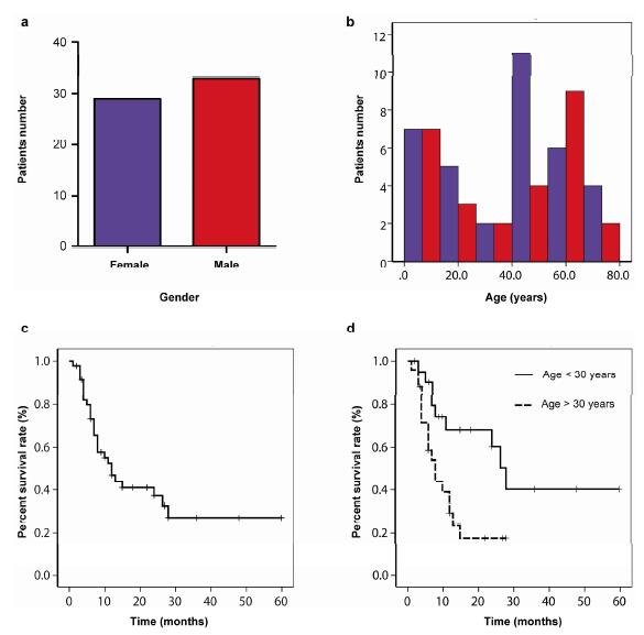
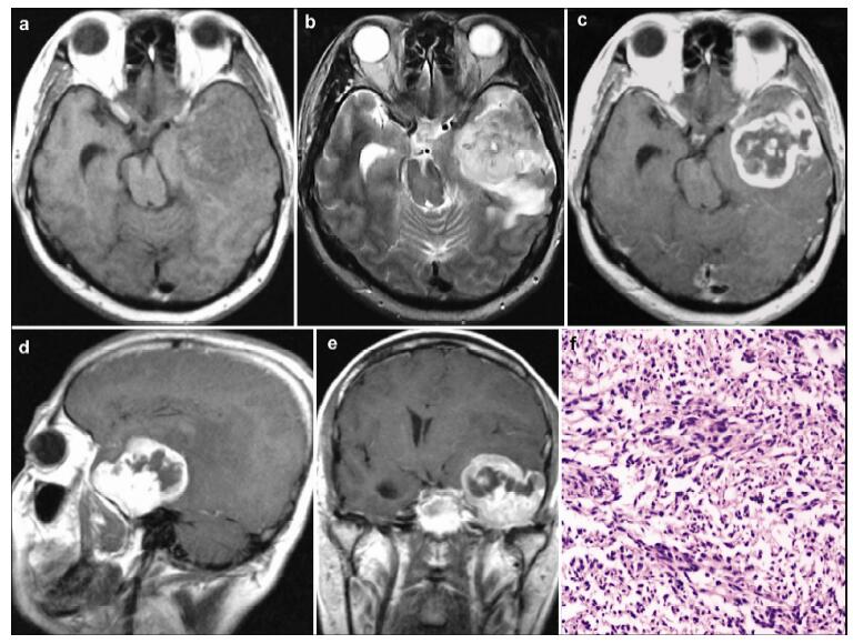
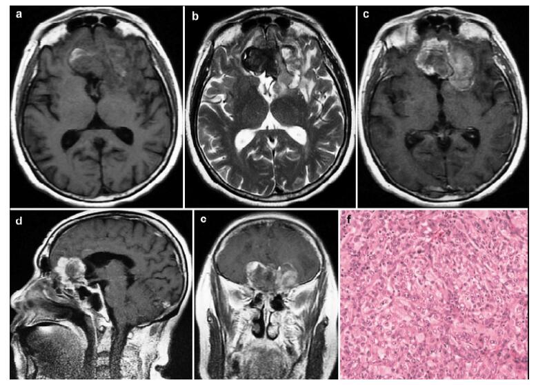
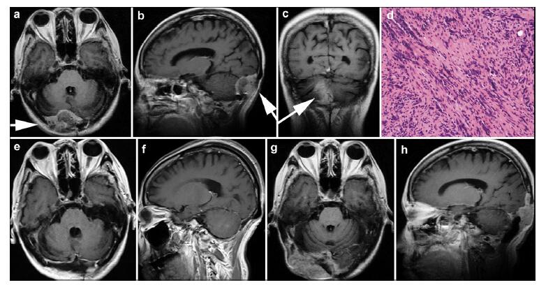
Abstract Objective: Primary intracranial malignant fibrous histiocytoma (MFH) is rare. We describe the detailed clinical features of 8 cases and fully review the literature to evaluate several prognostic factors. Methods: Eight patients with pathologically confirmed primary intracranial MFH were retrospectively reviewed. We searched PubMed for relevant articles with the term "intracranial malignant fibrous histiocytoma". Results: Of the 8 patients, 4 were men and 4 were women. Three patients had received previous radiotherapy. The age of the patients ranged from 19 to 69 years, with a median age of 48 years. Most tumors could be totally resected; and only 1 tumor was subtotally resected. Six patients received postoperative radiotherapy and 3 patients received postoperative chemotherapy. Most patients died within the first year after surgery; and only 1 patient was still alive on the date of the last follow-up. We reviewed the literature and included a total of 46 patients in the Kaplan-Meier survival analysis. Young patients (less than 30 years old) seemed to have a better prognosis and survival rate than older patients (more than 30 years old) (log-rank test, P=0.008). However, sex (P=0.675), extent of resection (P=0.934), postoperative radiotherapy (P=0.592), and postoperative chemotherapy (P=0.424) did not affect patient prognosis. Conclusions: The prognosis of MFH is usually poor, and most patients die within the first year after surgery. Younger MFH patients (less than 30 years old) seem to have a better prognosis and improved survival compared to older patients.
|
|
Received: 10 July 2016
Published: 30 September 2016
|
|
Corresponding Authors:
Pinan Liu, E-mail:pinanliu@ccmu.edu.cn
E-mail: pinanliu@ccmu.edu.cn
|
|
Cite this article:
Peng Li, Qiangyi Zhou, Zhijun Yang, Zhenmin Wang, Shiwei Li, Xingchao Wang, Bo Wang, Fu Zhao, Pinan Liu. Clinical features and prognostic factors of primary intracranial malignant fibrous histiocytoma: A report of 8 cases and a literature review. Translational Neuroscience and Clinics, 2016, 2(3): 155-164.
URL:
http://tnc.tsinghuajournals.com/10.18679/CN11-6030/R.2016.024 OR http://tnc.tsinghuajournals.com/Y2016/V2/I3/155
|
|
|
| [1] |
Pimentel J, Fernandes A, Távora L, Miguéns J, Lobo Antunes J. Benign isolated fibrohistiocytic tumor arising from the central nervous system. Considerations about two cases. Clin Neuropathol 2002, 21(3):93-98.<br />
|
| [2] |
Deb P, Singh V, Dutta V, Bhatoe HS, Chandran VM. Primary intracranial benign fibrous histiocytoma:Report of an unusual case. J Cancer Res Ther 2014, 10(1):200-202.<br />
|
| [3] |
Moliterno JA, Sood S, Zambrano E, Kim JH, Piepmeier JM, Baehring JM. Intracranial benign fibrous histiocytomas:A case report and review. J Neurooncol 2009, 92(2):203-209.<br />
|
| [4] |
Hayami J, Kurokawa I, Hashimoto K, Kusumoto K. Malignant fibrous histiocytoma of scalp with intracranial invasion 20 years after postoperative irradiation. J Craniofac Surg 2003, 14(1):74-77.<br />
|
| [5] |
Hamlat A, Adn M, Caulet-Maugendre S, Guegan Y. Cerebellar malignant fibrous histiocytoma:Case report and literature review. Neurosurgery 2004, 54(3):745-751.<br />
|
| [6] |
Gelabert-González M, Fernández-Villa JM, Reyes-Santías R. Histiocitoma fibroso maligno de duramadre. Neurocirugía 2003, 14(3):235-239.<br />
|
| [7] |
Broniscer A, Ke W, Fuller CE, Wu J, Gajjar A, Kun LE. Second neoplasms in pediatric patients with primary central nervous system tumors:The St. Jude Children's Research Hospital experience. Cancer 2004, 100(10):2246-2252.<br />
|
| [8] |
Mitsuhashi T, Watanabe M, Ohara Y, Hatashita S, Ueno H. Multifocal primary intracerebral malignant fibrous histiocytoma-Case report. Neurol Med Chir (Tokyo) 2004, 44(5):249-254.<br />
|
| [9] |
Maruno M, Ghulam Muhammad AKM, Taguchi J, Suzuki T, Wada K, Isaka T, Yoshimine T. Giant cell type of primary intracranial malignant fibrous histiocytoma:A case report. Brain Tumor Pathol 2006, 23(1):65-70.<br />
|
| [10] |
Harries AM, Mitchell R. Haemorrhagic cerebellar fibrous histiocytoma:Case report and literature review. Br J Neurosurg 2011, 25(1):120-121.<br />
|
| [11] |
Sarrami AH, Setareh M, Afshar-Moghaddam N, Izadinejad M, Saadatnia M. A case of intracranial malignant fibrous histiocytoma. J Res Med Sci 2011, 16(7):968-973.<br />
|
| [12] |
Gelincik I. Cerebellar malignant fibrous histiocytoma. Indian J Pathol Microbiol 2012, 55(3):402-405.<br />
|
| [13] |
Ozdemir M, Ozgural O, Bozkurt M, Torun FM, Heper AO, Tuna H. Primary intracerebral malignant fibrous histiocytoma mimicking a meningioma. Turk Neurosurg 2012, 22(4):475-477.<br />
|
| [14] |
Grahovac G, Chudy D, Heinrich Z, Zarkovic K. Implantation metastasis of malignant fibrous histiocytoma along the stereotactic biopsy tract. Clin Neurol Neurosurg 2013, 115(7):1160-1161.<br />
|
| [15] |
Yoo RE, Choi SH, Park SH, Jung HW, Kim JH, Sohn CH, Chang KH. Primary intracerebral malignant fibrous histio-cytoma:CT, MRI, and PET-CT findings. J Neuroimaging 2013, 23(1):141-144.<br />
|
| [16] |
Kurosaki M, Kambe A, Ishibashi M, Watanabe T, Horie Y. A case report of sarcoma of the sella caused by postoperative radiotherapy for a prolactin-producing pituitary adenoma. Brain Tumor Pathol 2014, 31(3):187-191.<br />
|
| [17] |
Özhan S, Tali ET, I?ik S, Saygili MR, Baykaner K. Haematoma-like primary intracranial malignant fibrous histiocytoma in a 5-year-old girl. Neuroradiology 1999, 41(7):523-525.<br />
|
| [18] |
Fritz MA, Sade B, Bauer TW, Wood BG, Lee JH. Benign fibrous histiocytoma of the pterygopalatine fossa with intracranial extension. Acta Neurochir 2006, 148(1):73-76.<br />
|
| [19] |
Graber JJ, Nayar A, Zagzag D. Metastatic cerebral malignant fibrous histiocytoma masquerading as neurocysticercosis. J Neurooncol 2011, 105(2):437-439.<br />
|
| [20] |
Schrader B, Holland BR, Friedrichsen C. Rare case of a primary malignant fibrous histiocytoma of the brain. Neuroradiology 1989, 31(2):177-179.<br />
|
| [21] |
Mahore A, Ramdasi R, Dange N, Epari S. Malignant fibrous histiocytoma of the skull base:A neurosurgical nuance. Asian J Neurosurg 2015, 10(2):135-138.<br />
|
| [22] |
Wang J, Zhong WM, Xu YH, Feng L, Li Y, Dong B. A primary malignant fibrous histiocytoma of the scalp and intracranial tumor bleeding:A case report. J Med Case Rep 2014, 8:50.<br />
|
| [23] |
Matsuura S, Takagi T, Tan EC, Mizuno S, Imagunbai N, Hasegawa R. Malignant fibrous histiocytoma of the occipital bone with intracranial extension-Case report. Neurol Med Chir (Tokyo) 1991, 31(4):219-222.<br />
|
| [24] |
Wu TH, Shih CW, Huang JS, Wang CH, Yeh KY. Unusual hematogenous brain metastasis in malignant fibrous histio-cytoma of the maxillary sinus. Int J Clin Oncol 2012, 17(1):69-74.<br />
|
| [25] |
Itoyama Y, Nagahiro S, Seto H, Sueyoshi N, Kuratsu JI, Ushio Y. Brain metastasis from malignant fibrous histiocytoma of the heart:Case report. Neurosurgery 1990, 26(4):692-695.<br />
|
| [26] |
Gonzalez-Vitale JC, Slavin RE, McQueen JD. Radiation-induced intracranial malignant fibrous histiocytoma. Cancer 1976, 37(6):2960-2963.<br />
|
| [27] |
Baehring JM, Alemohammed S, Croul SE. Malignant fibrous histiocytoma presenting as an intraventricular mass five years after incidental detection of a mass lesion. J Neurooncol 2001, 52(2):157-160.<br />
|
| [28] |
Hirato J, Nakazato Y, Sasaki A, Yokota M, Nojiri K, Toyoda O, Nakajima H. Intracranial malignant fibrous histiocytoma:Characterization of GFAP-positive cells in the tumor. Clin Neuropathol 1994, 13(6):315-322.<br />
|
| [29] |
Amendola BE, Amendola MA, McClatchey KD. Radiation-induced malignant fibrous histiocytoma:A report of five cases including two occurring post whole brain irradiation. Cancer Invest 1985, 3(6):507-513.<br />
|
| [30] |
Martinez-Salazar A, Supler M, Rojiani AM. Primary intracerebral malignant fibrous histiocytoma:Immunohisto-chemical findings and etiopathogenetic considerations. Mod Pathol 1997, 10(2):149-154.<br />
|
| [31] |
Paulus W, Peiffer J, Grote E. Intracerebral malignant fibrous histiocytoma at site of a previously excised low grade glioma. Acta Neurochir (Wien) 1989, 99(3-4):161-165.<br />
|
| [32] |
Bora H, Oztürk B, Akmansu M, Yenidunya S, Egehan I. Intracerebral malignant fibrous histiocytoma in a 5-year-old girl. Radiat Med 1999, 17(5):355-358.<br />
|
| [33] |
Kalyanaraman UP, Taraska JJ, Fierer JA, Elwood PW. Malignant fibrous histiocytoma of the meninges. Histological, ultrastructural, and immunocytochemical studies. J Neurosurg 1981, 55(6):957-962.<br />
|
| [34] |
Akimoto J, Takeda Y, Hasue M, Ito H, Kiguchi E. Primary meningeal malignant fibrous histiocytoma with cerebrospinal dissemination and pulmonary metastasis. Acta Neurochir (Wien) 1998, 140(11):1191-1196.<br />
|
| [35] |
Ho YS, Wei CH, Tsai MD, Wai YY. Intracerebral malignant fibrous histiocytoma:Case report and review of the literature. Neurosurgery 1992, 31(3):567-570.<br />
|
| [36] |
Berry AD 3rd, Reintjes SL, Kepes JJ. Intracranial malignant fibrous histiocytoma with abscess-like tumor necrosis. Case report. J Neurosurg 1988, 69(5):780-784.<br />
|
| [37] |
Kepes JJ. "Xanthomathous" lesions of the central nervous system:Definition, classification and some recent observations. In Progress in Neuropathology Vol. 4. Zimmerman HMN, Ed. New York:Raven, 1979, pp 179-213.<br />
|
| [38] |
Akai T, Yamamoto K, Iida T, Iizuka H, Nojima T. Malignant fibrous histiocytoma in the craniocervical junction presenting with severe occipitalgia. Brain Tumor Pathol 2006, 23(2):101-105.<br />
|
| [39] |
Le Doussal V, Coindre JM, Leroux A, Hacene K, Terrier P, Bui NB, Bonichon F, Collin F, Mandard AM, Contesso G. Prognostic factors for patients with localized primary malignant fibrous histiocytoma:A multicenter study of 216 patients with multivariate analysis. Cancer 1996, 77(9):1823-1830.<br />
|
| [40] |
Fujimura N, Sugita Y, Hirohata M, Naohisa M, Terasaki M, Tokutmi T, Shigemori M. Primary intracerebral malignant fibrous histiocytoma in a child. Pediatr Neurosurg 2002, 37(5):271-274.<br />
|
| [41] |
Pople IK, Harding B. Primary intracranial malignant fibrous histiocytoma in a 5-year-old boy:Case report. Br J Neurosurg 1991, 5(5):509-513.<br />
|
| [42] |
Chhabra R, Gupta SK, Manjunath Prasad KS, Gupta D, Vasishta RK, Sharma RK, Khosla VK. Calvarial malignant fibrous histiocytoma. Neurol India 2004, 52(3):387-390.
|
|
Viewed |
|
|
|
Full text
|
|
|
|
|
Abstract
|
|
|
|
|
Cited |
|
|
|
|
| |
Shared |
|
|
|
|
| |
Discussed |
|
|
|
|