| Short Communications |
|


|
|
|
| Complete resection of cavernous malformations in the hypothalamus: A case report and review of the literature |
| Xingchao Wang1, Zhenmin Wang1, Zhixian Gao1, Pinan Liu1,2 |
1 Department of Neurosurgery, Beijing Tiantan Hospital, Capital Medical University, Beijing 100050, China;
2 Department of Neural Reconstruction, Beijing Neurosurgery Institute, Capital Medical University, Beijing 100050, China |
|
|
|
|
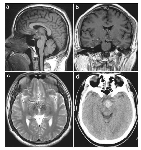
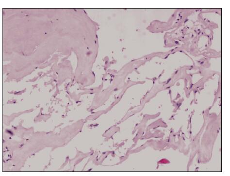
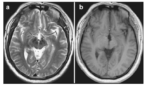
Abstract Objective: Cavernous malformation (CM) originating from the hypothalamus is extremely rare and the deep location presents a challenge for its neurosurgical management. We report such a case to better understand its clinical features. Methods and Results: A 40-year-old male patient presented with impaired vision in the left eye. Magnetic resonance imaging (MRI) revealed a regularly shaped round lesion located in the suprasellar cistern, and a clinical diagnosis of hypothalamic CM was made. Complete microsurgical excision was performed via a right pterional craniotomy. The patient showed good recovery with no further visual acuity or field deficits postoperatively. No CM recurrence or rebleeding was seen on follow-up MRI scans performed over the course of two years. Conclusions: For patients with cavernous malformation in the hypothalamus, accurate preoperative diagnosis with complete surgical removal by an appropriate surgical approach can contribute to satisfactory outcomes.
|
|
Received: 05 July 2016
Published: 30 September 2016
|
|
Corresponding Authors:
Pinan Liu, E-mail:pinanliu@ccmu.edu.cn;Zhixian Gao, E-mail:elunlun0555@sina.com
E-mail: pinanliu@ccmu.edu.cn;elunlun0555@sina.com
|
|
|
| [1] |
Batra S, Lin D, Recinos PF, Zhang J, Rigamonti D. Cavernous malformations:Natural history, diagnosis and treatment. Nat Rev Neurol 2009, 5(12):659-670.<br />
|
| [2] |
Hassler W, Zentner J, Wilhelm H. Cavernous angiomas of the anterior visual pathways. J Clin Neuroophthalmol 1989, 9(3):160-164.<br />
|
| [3] |
Mizutani T, Goldberg HI, Kerson LA, Murtagh F. Cavernous hemangioma in the diencephalon. Arch Neurol 1981, 38(6):379-382.<br />
|
| [4] |
Reyns N, Assaker R, Louis E, Lejeune JP. Intraventricular cavernomas:Three cases and review of the literature. Neurosurgery 1999, 44(3):648-654.<br />
|
| [5] |
Samii M, Eghbal R, Carvalho GA, Matthies C. Surgical management of brainstem cavernomas. J Neurosurg 2001, 95(5):825-832.<br />
|
| [6] |
Abou-Al-Shaar H, Bahatheq A, Takroni R, Al-Thubaiti I. Optic chiasmal cavernous angioma:A rare suprasellar vascular malformation. Surg Neurol Int 2016, 7(Suppl 18):S523-S526.<br />
|
| [7] |
Liu JK, Lu Y, Raslan AM, Gultekin SH, Delashaw JB Jr. Cavernous malformations of the optic pathway and hy-pothalamus:Analysis of 65 cases in the literature. Neurosurg Focus 2010, 29(3):E17.<br />
|
| [8] |
Mizoi K, Yoshimoto T, Suzuki J. Clinical analysis of ten cases with surgically treated brain stem cavernous angiomas. Tohoku J Exp Med 1992, 166(2):259-267.<br />
|
| [9] |
Kurokawa Y, Abiko S, Ikeda N, Ideguchi M, Okamura T. Surgical strategy for cavernous angioma in hypothalamus. J Clin Neurosci 2001, 8 Suppl1:106-108.<br />
|
| [10] |
Katayama Y, Tsubokawa T, Maeda T, Yamamoto T. Surgical management of cavernous malformations of the third ventricle. J Neurosurg 1994, 80(1):64-72.<br />
|
| [11] |
Rheinboldt M, Blase J. Exophytic hypothalamic cavernous malformation mimicking an extra-axial suprasellar mass. Emerg Radiol 2011, 18(4):363-367.<br />
|
| [12] |
Gross BA, Du R. Cerebral cavernous malformations:Natural history and clinical management. Expert Rev Neurother 2015, 15(7):771-777.<br />
|
| [13] |
Simard JM, Garcia-Bengochea F, Ballinger WE Jr, Mickle JP, Quisling RG. Cavernous angioma:A review of 126 collected and 12 new clinical cases. Neurosurgery 1986, 18(2):162-172.<br />
|
| [14] |
Hempelmann RG, Mater E, Schröder F, Schön R. Complete resection of a cavernous haemangioma of the optic nerve, the chiasm, and the optic tract. Acta Neurochir (Wien) 2007, 149(7):699-703; discussion 703.<br />
|
| [15] |
Wang CH, Lin SM, Chen Y, Tseng SH. Multiple deep-seated cavernomas in the third ventricle, hypothalamus and thalamus. Acta Neurochir (Wien) 2003, 145(6):505-508.<br />
|
| [16] |
Ogawa Y, Tominaga T. Sellar and parasellar tumor removal without discontinuing antithrombotic therapy. J Neurosurg 2015, 123(3):794-798.<br />
|
| [17] |
Hasegawa H, Bitoh S, Koshino K, Obashi J, Kobayashi Y, Kobayashi M, Wakasugi C. Mixed cavernous angioma and glioma (angioglioma) in the hypothalamus-Case report. Neurol Med Chir (Tokyo) 1995, 35(4):238-242.<br />
|
| [18] |
Robinson JR, Awad IA, Little JR. Natural history of the cavernous angioma. J Neurosurg 1991, 75(5):709-714.<br />
|
| [19] |
Porter RW, Detwiler PW, Spetzler RF, Lawton MT, Baskin JJ, Derksen PT, Zabramski JM. Cavernous malformations of the brainstem:Experience with 100 patients. J Neurosurg 1999, 90(1):50-58.<br />
|
| [20] |
Lehner M, Fellner FA, Wurm G. Cavernous haemangiomas of the anterior visual pathways. Short review on occasion of an exceptional case. Acta Neurochir (Wien) 2006, 148(5):571-578.
|
|
Viewed |
|
|
|
Full text
|
|
|
|
|
Abstract
|
|
|
|
|
Cited |
|
|
|
|
| |
Shared |
|
|
|
|
| |
Discussed |
|
|
|
|